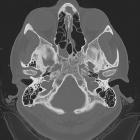
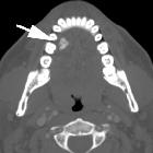
arrested pneumatization of skull base


Arrested pneumatization of the skull base is an anatomical variant that most commonly occurs in association with the sphenoid sinus. It is known that the sphenoid bones undergo early fatty marrow conversion antecedent to normal pneumatization. However, for unclear reasons, some individuals experience failure of pneumatization before respiratory mucosa has fully extended into sites of early fatty marrow conversion. These individuals are then left with persistent atypical fatty marrow adjacent to the sinus that persists into adulthood. If unrecognised, imaging features may at times create diagnostic difficulty in interpretation of skull base CT and MR scans.
Clinical presentation
Since it is a developmental abnormality, it usually manifests as an incidental finding. No specific clinical symptoms are attributed to this entity.
Pathology
The proper process of pneumatization of the skull base and paranasal sinuses start at the age of 4 months and develops through to young adulthood. Red bone marrow is being replaced by fatty marrow prior to pneumatization of the paranasal sinuses, including the sphenoid bone. The precise mechanisms underlying this process remain largely unclear. This bone marrow conversion precedes the invasion of epithelial cells to form the respiratory mucosa. When one of the steps described above is halted, no or reduced pneumatization of the sinus will occur.
Location
It usually occurs in regions of normal or accessory sphenoid sinus pneumatization including the basisphenoid bone, pterygoid processes and clivus. Contiguous involvement across multiple sphenoid subsites is common.
Radiographic features
Imaging consists of CT and MR studies involving the skull base. The non-expansile nature of the lesion is best evaluated at the inferior orbital fissure and Vidian canal, which are not displaced nor disrupted.
CT
Characteristic features on CT are the presence of:
- a non-expansile lesion with
- internal curvilinear calcifications and
- sclerotic margins
MRI
Hallmark of MR imaging is:
- the presence of internal fat and microcystic components
- the absence of any mass effect
- T1
- hyperintense fatty component
- hypointense microcystic and calcified components
- sclerotic border is hypointense
- no enhancement of the lesion after Gd administration
- T2: microcystic components are hyperintense on T2
Differential diagnosis
General differential considerations include:
- fibrous dysplasia
- ossifying fibroma
- chondrosarcoma
- osteomyelitis (around skull base)
- chordoma
- bone metastasis(es)
In contrast with arrested pneumatization, all of these conditions lack the presence of internal fat or usually show signs of mass effect on the surrounding structures.
Practical points
Arrested pneumatization can be diagnosed when a lesion fulfills the following criteria:
- the lesion must be located at a site of normal pneumatization or of recognized accessory pneumatization
- the lesion must be non-expansile with sclerotic, well-circumscribed margins
- the lesion should show fatty content
- on CT, internal curvilinear calcifications should be present
- any associated skull base foramina should retain a normal appearance
Siehe auch:

 Assoziationen und Differentialdiagnosen zu arrested pneumatization of skull base:


