Bronchoceles


Mucoid impaction, also referred to as mucus plugging, mucous plugging, bronchial mucocele or bronchocele formation, refers to airway filling by mucoid secretions and can be obstructive or non-obstructive. It is a common pathological finding in chest imaging.
Pathology
Etiology
Mucoid impaction may result from either obstructive or non-obstructive causes, although the latter does eventually obstruct the bronchi as well:
Non-obstructive
Non-obstructive causes are infectious or inflammatory in nature:
- cystic fibrosis: due to impaired ciliary movement and thick secretions
- asthma: due to increased mucus production
- allergic bronchopulmonary aspergillosis (ABPA) - can sometimes have high attenuation mucoid impaction
Obstructive
- congenital
- congenital bronchial atresia: most commonly affects the apicoposterior segment of the left upper lobe
- intralobar sequestration
- intrapulmonary bronchogenic cyst
- neoplastic
- other acquired conditions
- broncholithiasis
- tuberculous bronchostenosis
- foreign body aspiration
Radiographic features
Plain radiograph
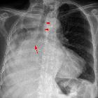
Mucoid impaction may appear as a branching tubular opacity that is distinct from the normal vascular shadows.This classic feature is the finger-in-glove sign, and is also seen on CT. It can sometimes prove difficult to diagnose on plain radiography.
CT
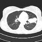
The appearance on CT can be variable, depending on where the mucous plugging occurs (i.e. central or peripheral airways).
Mucous plugs appear as fluid density material often located dependently within the bronchus, sometimes with internal gas.
The classic feature seen when plugged mucus extends along large branching airways is the finger-in-glove sign.
Several key additional features can also occur:
- distal lung collapse: from collateral air drift; this has not yet developed in infants and young children, which is why the distal lung is rather hyperlucent in this population (e.g. congenital bronchial atresia, foreign body aspiration)
- distal airway dilatation
- double artery sign
Treatment and prognosis
Complications
Prolonged mucous plugging can lead to bronchial dilatation and bronchiectasis.
Differential diagnosis
- endobronchial blood or blood clot appears similar to mucous plugging on CT
- in some situations, there can be overlap between some endobronchial tumors per se (rather than with associated mucoid impaction)
Siehe auch:
- Bronchiektasen
- Lipom
- Lungenkarzinom
- Bronchogene Zyste
- arteriovenöse Malformationen der Lunge
- Allergische bronchopulmonale Aspergillose
- Hamartom
- zystische Fibrose
- Bronchialatresie
- bronchiales Karzinoid
- mucoid impaction - cystic fibrosis
- hyperdense Mukusimpaktion
- Finger in Handschuh Bild
- sequestration
und weiter:


 Assoziationen und Differentialdiagnosen zu Bronchozele: