bronchopulmonary dysplasia


Bronchopulmonary dysplasia (BPD) refers to late pathological lung changes that develop in infants after several weeks on prolonged ventilation.
Terminology
BPD and chronic lung disease of prematurity (CLDP) have often been used interchangeably to describe the condition post-treatment of premature infants for respiratory distress syndrome. However, some suggest that there are different underlying pathogeneses and that CLDP encompasses other conditions besides BPD .
Pathology
It is the result of a paradoxical combination of hypoxia and oxygen toxicity. There is initial capillary wall damage, interstitial fluid seepage and ensuing pulmonary edema, which is followed by loss of ciliated epithelium and bronchiolar mucosal necrosis. Areas of both hyperexpansion and atelectasis are seen. This is followed by eosinophilic exudate and squamous metaplasia and may ultimately lead to interstitial fibrosis/fibro-proliferative bronchiolitis.
Radiographic features
Plain radiograph
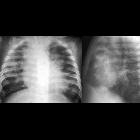
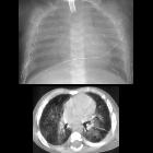
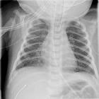
- ill-defined reticular markings with interspersed rounded lucent areas diffusely involving hyperinflated lungs
- the lungs may have relatively normal AP diameter on the lateral film
- presence of cardiomegaly may indicate the development of pulmonary hypertension
- in chronic cases, the lateral film may show a much narrower AP diameter compared with the chest width on the frontal film
CT
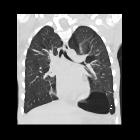
- mosaic lung parenchymal pattern with areas of low attenuation and focal air trapping on expiratory HRCT (considered the most sensitive finding for predicting severity)
- bronchial wall thickening (considered the most frequent finding)
- small subpleural triangular/linear opacities
Bronchiectatic changes are usually not considered a feature .
Treatment and prognosis
Infants who survive neonatal bronchopulmonary dysplasia often show a slow but continuous improvement in respiratory status. Young adult survivors who have had moderate and severe bronchopulmonary dysplasia may have residual functional and characteristic structural pulmonary abnormalities; of these, the most notable is pulmonary emphysema .
Differential diagnosis
General imaging differential considerations include:
- pulmonary interstitial emphysema (has an acute course)
- neonatal pneumonia
- Wilson-Mikity syndrome
See also
Siehe auch:
und weiter:

 Assoziationen und Differentialdiagnosen zu Bronchopulmonale Dysplasie:

