congenital megaureter


A congenital (primary) megaureter encompasses causes of an enlarged ureter which are intrinsic to the ureter, rather than as a result of a more distal abnormality; e.g. bladder, urethra (see secondary megaureter). It includes:
- obstructed primary megaureter
- refluxing primary megaureter
- although vesicoureteric reflux (VUR) is a cause of primary congenital megaureter it is usually considered separately
- non-refluxing unobstructed primary megaureter
Clinical presentation
In all three types of megaureter, patients are often asymptomatic. Symptoms, when present, are usually arise from complications due to urinary stasis (e.g. urinary sepsis and nephrolithiasis).
Congenital primary megaureter is sometimes associated with:
- congenital megacalyces
- ipsilateral renal dysplasia
Pathology
Obstructive primary megaureter
Obstructive primary megaureter is related to a distal adynamic segment with proximal dilatation and is a common cause of obstructive uropathy in children . It is analogous to esophageal achalasia or colonic Hirschsprung disease although a lack of ganglion cells within the wall of the ureter has not been proven to be the cause .
Refluxing primary megaureter
Refluxing primary megaureter is a result of an abnormal vesicoureteric junction, which impedes the normal anti-reflux mechanisms. This can be due to a short vertical intramural segment, congenital paraureteric diverticulum, ureterocele with or without associated duplicated collecting system, etc.
It is relatively common and usually considered separately (see vesicoureteric reflux (VUR)).
Non-refluxing unobstructed primary megaureter
This is thought to be the most common cause of primary megaureter in neonates, and even though the vesicoureteric junction is normal, with no evidence of reflux or obstruction the ureter is enlarged. The reason for this is unknown.
Radiographic features
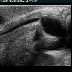
In all three types the ureter is enlarged (>7 mm) sometimes markedly so. On all modalities able to visualize the ureter (CT, US, MRI, IVP) it appears as a tubular structure usually posterior to the bladder .
In obstructive primary megaureter the ureter tapers to a short segment of normal caliber or narrowed distal ureter, usually just above the vesicoureteric junction (VUJ). The distal ureter above this narrowed segment is most dilated (similar to achalasia). There is associated hydronephrosis, and active peristaltic waves can be seen on ultrasound.
In refluxing primary megaureter, vesicoureteric reflux is demonstrated (see vesicoureteric reflux (VUR) )
In non-refluxing unobstructed primary megaureter, there is absent or only a minor degree of hydronephrosis. Although rare, a congenital megaureter may co-exist with congenital megacalyces , making the assessment of hydronephrosis more difficult.
Treatment and prognosis
Usually asymptomatic and requiring no treatment. If complications occur or the degree of obstruction is marked then, reimplantation following resection of the aganglionic segment may be performed.
Differential diagnosis
Differential diagnostic considerations include:
- vesicoureteric reflux disease (VUR): although a cause of primary congenital megaureter it is usually considered separately as prognosis and treatment, depending on the degree of reflux, is different
- secondary megaureter
- posterior urethral valves: often bilateral hydroureter/hydronephrosis
- ureteral diverticulum
- urolithiasis
- duplex collecting system with reflux into lower pole moiety
Siehe auch:
und weiter:


 Assoziationen und Differentialdiagnosen zu kongenitaler Megaureter: