desmoplastic fibroma


Desmoplastic fibromas are extremely rare bone tumors that do not metastasize but may be locally aggressive. They are considered to be a bony counterpart of soft tissue desmoid tumors and are histologically identical.
Epidemiology
Desmoplastic fibroma of bone is rare and mostly found in young adults and adolescents. It accounts for less than 0.1% of all bone tumors . There is no sex predilection.
Clinical presentation
Usual complaints are pain or deformity. Some patients might also present with a fracture or the tumor might be found incidentally .
Complications
If left untreated it can lead to the following :
- pathologic fracture
- transition to osteosarcoma (extremely rare)
Pathology
Desmoplastic fibroma of bone is a tumor made up of spindle cells resembling desmoid-type fibromatosis and features an infiltrative growth pattern .
Etiology
The etiology of desmoplastic fibroma of bone is unknown.
Location
Most frequent locations of desmoplastic fibroma of bone include :
- mandible
- long bones (femur, radius, tibia)
- usually located in the metaphysics or diametaphysis
- pelvic bones
Macroscopic appearance
Macroscopically desmoplastic fibroma of bone usually appears as a whorled, creamy-colored tumor due to high collagen content featuring a firm consistency .
Microscopic appearance
Microscopic features of desmoplastic fibroma of bone include the following :
- low cellularity consistent of bland spindle cells in a collagenous matrix
- scant mitoses
- no necroses
- main cell types include fibroblasts, myofibroblasts, and undifferentiated mesenchymal cells
Immunohistochemistry
Immunohistochemistry stains can be positive for β-catenin.
Radiographic features
General imaging features of desmoplastic fibroma of bone comprise the following :
- usually well-defined or partly well-defined
- narrow transition zone
- lobulated form
- expansile growth
- sometimes with cortical bone destruction and extension into the surrounding tissues
Plain radiograph
On plain radiographs desmoplastic fibroma of bone will usually show the following characteristics :
- osteolytic or mixed osteolytic/mildly sclerotic matrix
- well-defined or partly well-defined margins
- possible endosteal scalloping or cortical breakthrough
- geographic pattern of bone destruction
- often non-sclerotic margins
- internal pseudotrabeculation: >90%
- no matrix mineralization
- widening of the host bone from gradual apposition of periosteal new bone formation: ~90%
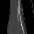
CT
CT will show a soft tissue density mass and will show cortical arrosion in a high percentage possibly associated with infiltration of the surrounding tissues .
MRI
In addition to the general imaging features, MRI shines in depicting soft tissue expansion . In addition to the solid tumor component, there might be cystic changes present.
Signal characteristics are usually as follows:
- T1: low signal intensity (isointense or hypointense to muscle)
- T2: background intermediate to high signal with intrinsic low to intermediate intensity foci within
- T1 C+ (Gd): heterogeneous enhancement
Nuclear medicine
Bone scintigraphy/FDG PET
Desmoplastic fibroma of bone might show increased uptake .
Radiology report
The radiological report should include a description of the following:
- form and location
- tumor margins and transition zone
- endosteal scalloping
- cortical breakthrough
- infiltration of the surrounding tissues
Treatment and prognosis
Despite being benign it is a still locally aggressive tumor. En bloc resection is the current treatment of choice.
Due to its rarity prognosis is difficult, but it is considered a slow-growing progressive tumor, which shows a local aggressive behavior .
History and etymology
Initially described by HL Jaffe in 1958 .
Differential diagnosis
Conditions which can mimic the presentation and/or the appearance of desmoplastic fibroma of bone include:
- osteosarcoma
- osteoblastoma
- non-ossifying fibroma
- chondromyxoid fibroma
- brown tumor
- giant cell tumor of bone (GCT)
See also
Siehe auch:
und weiter:


 Assoziationen und Differentialdiagnosen zu desmoplastisches Fibrom:


