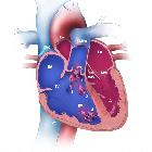
ectopia cordis


Ectopia cordis is an extremely rare congenital malformation where the heart is located partially or totally outside the thoracic cavity. The four main ectopic positions are:
- adjacent to the thorax: ~60%
- abdominal: 15-30%
- thoracoabdominal: 7-18%
- cervical: ~3%
Epidemiology
The incidence is 5.5 to 7.9 per 1,000,000 live births .
Pathology
It results from the failure of migration of lateral mesoderm into the midline. Some forms may be the outcome of amniotic band syndrome .
Ectopia cordis may occur as an isolated malformation or it may be associated with a larger category of ventral body wall defects that affect the thorax, abdomen, or both.
Associations
Individual associations
Ventricular septal defect and tetralogy of Fallot are the most common intracardiac defects. Omphalocele is the most common abdominal wall defect.
Syndromic associations
A well-known association is pentalogy of Cantrell which comprises:
- ectopia cordis
- omphalocele (typically supraumbilical)
- congenital diaphragmatic hernia
- sternal cleft
- congenital heart disease
Radiographic features
Antenatal ultrasound
When in isolation, the heart is seen in the amniotic cavity with a thoracic wall defect. If in association with pentalogy of Cantrell, it may seen within an omphalocele .
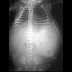
Plain radiograph
Imaging clues on frontal chest radiographs include:
- abnormal cardiac position and configuration
- air lucency may surround the ectopic heart
- sternal defect is often present
- wide separation of the sternal ends of the clavicles
- widening of the superior mediastinum
The lateral view may confirm the extrathoracic location of the heart.
CT/MRI
Cross-sectional modalities allow for the best visualization of ectopia cordis, and is required for any surgical planning.
Treatment and prognosis
Although surgical correction may be attempted , the prognosis is generally poor and depends on the severity of intracardiac malformations and the presence of associated abnormalities. Most infants are stillborn or die within the first hours or days of life.
Siehe auch:
- Herzfehler
- Fallot'sche Tetralogie
- Omphalozele
- Herz Anatomie
- kongenitale Zwerchfellhernie
- Ventrikelseptumdefekt
- Cantrell’sche Pentalogie
- Fissura sterni congenita
- cardiac herniation
und weiter:


 Assoziationen und Differentialdiagnosen zu Herzektopie: