Foramen jugulare

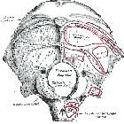
The jugular foramen courses anteriorly, laterally, and inferiorly as it insinuates itself between the petrous temporal bone and the occipital bone.
Gross anatomy
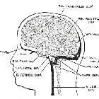
The jugular foramen is usually described as being divided into two parts by a fibrous or bony septum, called the jugular spine, into:
- the pars nervosa: anteromedial and smaller
- the pars vascularis: posterolateral and larger
Pars nervosa
The pars nervosa is the anteromedial portion of the jugular foramen and is smaller than the larger, posterolateral pars vascularis. It contains:
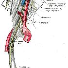
The inferior petrosal sinus drains the cavernous sinus and courses in the petro-occipital fissure adjacent to the clivus prior to its exit through the pars nervosa and subsequent drainage into the internal jugular vein beneath the foramen.
The glossopharyngeal nerve yields a tympanic branch (Jacobson nerve) which reaches and supplies the middle ear along with the inferior tympanic artery via the inferior tympanic canaliculus which is occasionally seen at CT in cross-section at the level of the caroticojugular spine.
Pars vascularis
The pars vascularis is the posterolateral portion of the jugular foramen and is larger than the smaller anteromedial portion termed the pars nervosa. It contains:
- jugular bulb: a venous expansion sitting in the jugular fossa, between the endocranial sigmoid sinus and the exocranial jugular vein
- vagus nerve (CN X)
- spinal accessory nerve (CN XI)
The vagus nerve yields an auricular branch (Arnold nerve) via the mastoid canaliculus on the lateral wall of the foramen adjacent to the mastoid segment of the facial nerve.
Alternative division into 3 parts
In this system the foramen is divided into 3 parts:
The intrajugular part is positioned between the sigmoid and petrosal parts, and is the site of bony prominences called the intrajugular processes on the opposing surfaces of the temporal and occipital bones.
Radiographic features
The size of the normal jugular foramen is remarkably variable and asymmetrical. However, the normal foramen will always be well-corticated. Therefore, the bony margin (and not the size) of the foramen should guide diagnostic evaluation.
CT
CT appearance of the foramen depends upon the level and angulation of the scan. The foramen has been described as having the shape of a sitting duck on the highest axial cuts.
Siehe auch:
- Bulbus-venae-jugularis-Hochstand
- Sinus sigmoideus
- Paragangliom
- Nervus glossopharyngeus
- inferior petrosal sinus
- Nervus vagus
- Foramina der Schädelbasis
- Nervus accessorius
- dehiszenter Bulbus jugularis
- Arteria meningea posterior
- Vernet's syndrome
- Bulbus superior venae jugularis internae
- Arnold nerve
- sitting duck
- spinal accessory cranial nerve (CN XI)
und weiter:


 Assoziationen und Differentialdiagnosen zu Foramen jugulare: