gliomas

The
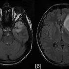
importance of fMRI and DTI in the presurgical planning of multicentric low-grade gliomas: a case report.. Axial FLAIR (Fluid Attenuated Inversion Recovery) images (A, B) reveal the high signal intensity of the two lesions located at the right temporal lobe and the left frontotemporal lobe regions respectively accompanied with ring-like oedema.

The
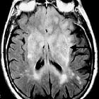
importance of fMRI and DTI in the presurgical planning of multicentric low-grade gliomas: a case report.. Axial FA colour-mapping images (A-D). The tumours, located at the right temporal and left frontotemporal regions, are visualized as a well-circumscribed bluish area, which is characteristic of low fractional anisotropy.
Gliom
gliomas
Siehe auch:
- Gliomatosis cerebri
- Glioblastoma multiforme
- butterfly glioma
- WHO-Klassifikation der Tumoren des zentralen Nervensystems
- Tectumgliom
- Opticusgliom
- zerebelläres Gliom
- high grade gliomas
- niedriggradiges Gliom
- multizentrisches niedriggradiges Gliom
- multimodality imaging in glioma recurrence
- Hirnstammgliom
und weiter:

 Assoziationen und Differentialdiagnosen zu Gliom:


