leiomyoma

Leiomyome des
Ösophagus: Links und Mitte: Fälle aus der Breischluckuntersuchung: Glatte Begrenzung der in das Lumen ragenden Raumforderung. Oberhalb ist das Lumen durch die peristaltische Kontraktion nicht mehr sichtbar (links). Der Winkel zur angrenzenden Lumenkontur ist stumpf (submuköse Lage). Rechts: In der CT kann die rundliche Konfiguration des Tumors oft erst in sagittalen oder koronaren Reformatierungen dargestellt werden.

Großes
Leiomyom des Magens in der Doppelkontrastuntersuchung. Glatt begrenzter Tumor ohne Schleimhautdestruktion; häufigster submuköser Tumor im Magen; meist Zufallsbefund; kann jedoch auch ulzerieren und Ursache einer oberen GI-Blutung sein! Differentialdiagnostisch kommen in Frage: Leiomyoblastom, -sarkom, Lipom (negative Dichtewerte in der CT!), Hämangiom, Neurofibrom und Granular-Zell-Tumor, ektoper Pankreasrest, akutes Ulkus des Antrums, Kompression von außen durch vergrößerte Nachbarorgane oder Metastasen in diesen (Leber, Milz, Pankreas, Niere)

Computertomographie:
Bild einer Lymphangioleiomyomatose bei Patientin mit multiplen Angiomyolipomen der Nieren: Dringender Verdacht auf Tuberöse Sklerose.

Calcified
uterine fibroid, as light calcification at paramedial right on pelvis minor (at left in image).


Leiomyoma of
the bladder in a 58-year-old woman. This mass is not vascularized on Doppler analysis.

Leiomyoma •
Angioleiomyoma - Ganzer Fall bei Radiopaedia

Management of
a giant retroperitoneal leiomyoma: a case report. A retroperitoneal tumor with a mass effect on the left kidney, the aorta, and the bowel. 1 = Tumor, 2 = left kidney, 3 = aorta, 4 = left kidney artery, 5 = iliac artery, 6 = pancreas

Imaging of
the oesophagus: beyond cancer. Leiomyoma. Esophagram in AP projection demonstrates a smooth, lobulated filling defect within the mid-thoracic oesophagus at the level of the carina. Notice the filling defect makes an acute angle to the oesophageal wall, suggesting that this lesion is intrinsic to the oesophagus
Leiomyoma is a benign smooth muscle (myometrial) tumor, most commonly found in the uterus.
Classification
Leiomyoma is classified by location:
- uterine leiomyoma
- cervical leiomyoma
- leiomyoma of the urinary bladder
- urethral leiomyoma
- solitary cutaneous leiomyoma
- vascular leiomyoma (angioleiomyoma)
- dartoic leiomyoma - arises from the dartos muscle of the nipple or genitals.
- esophageal leiomyoma
- leiomyoma of the jejunum
See also
- leiomyosarcoma
- angiolipoleiomyoma
- diffuse uterine leiomyomatosis
- diffuse peritoneal leiomyomatosis
- esophageal leiomyomatosis
- hereditary leiomyomatosis and renal cell carcinoma
- intravenous leiomyomatosis
- benign metastasizing leiomyoma
- parasitic leiomyoma
- extra-uterine pelvic leiomyoma
Siehe auch:
- Leiomyom des Magens
- Leiomyom Ösophagus
- diffuse uterine leiomyomatosis
- Leiomyom der Harnblase
- benign metastasising leiomyoma
- intra-mural uterine leiomyoma
- Leiomyom der Zervix uteri
- pelvine extrauterine Leiomyome
- hereditary leiomyomatosis and renal cell cancer (HLRCC)
- Leiomyomatose der Venen
- Leiomyomatosis peritonealis disseminata
- Leiomyom des Uterus mit zystischer Degeneration
- Leiomyom des Uterus mit hyaliner Degeneration
- gastrointestinal stromal tumors - leiomyoma/leiomyosarcoma
und weiter:


 Assoziationen und Differentialdiagnosen zu Leiomyom:
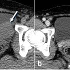


Leiomyom des
Uterus mit zystischer Degeneration