Lipome des Gastrointestinaltraktes


Gastrointestinal tract lipomas are not uncommon and can be found anywhere along the entire length of the gastrointestinal tract.
For a more specific discussion, please refer to the articles on:
Epidemiology
Gastrointestinal tract lipomas are most frequently encountered between the ages of 50 and 70 years .
Clinical presentation
The majority of lipomas are asymptomatic and found incidentally. As they can be pedunculated (see below) they occasionally present as the leading point of an intussusception. When large they may develop mucosal ulceration and present with iron deficiency anemia or positive fecal occult blood testing . Acute heavy bleeding is uncommon.
Pathology
Gastrointestinal lipomas, like lipomas elsewhere, are composed of mature adipocytes with an enveloping fibrous capsule .
The vast majority (90-95%) are submucosal, with only a small number subserosal, and can be sessile or pedunculated .
Location
- colon
- most common
- usually right-sided
- found in up to 0.25% of autopsies
- small intestine
- stomach: rare (see: gastric lipoma)
- esophagus: rare (see: esophageal lipoma)
Radiographic features
Fluoroscopy
Barium studies
Lipomas are usually submucosal or occasionally pedunculated. They usually have a very smooth surface, unless mucosal ulceration is present.
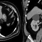
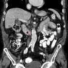
CT / MRI
On both CT and MRI lipomas are usually easy to diagnose on account of their density (-80 to -120 HU) / intensity following that of fat on all sequences. Lipomas are usually entirely of fat density without solid components. If a solid non-fat component is seen then the possibility of the mass representing a liposarcoma should be entertained, although these are exceedingly rare . Overlying ulceration may result in some non-fat density/intensity stranding near the mucosal surface.
Treatment and prognosis
As these are benign slow-growing lesions, and usually little doubt exists as to the diagnosis, no treatment is required. If symptomatic then local excision is sufficient .
Siehe auch:
- Lipom
- Invagination
- Kolonlipom
- Lipome des Ösophagus
- Duodenallipom
- Lipom des Magens
- gastrointestinale Lipomatose
- lipoma of the small intestine
- kolokolische Invagination ausgelöst durch Kolonlipom
- Invagination durch Lipom ausgelöst
- jejunal lipoma
und weiter:


 Assoziationen und Differentialdiagnosen zu Lipome des Gastrointestinaltraktes:


