normale intrakranielle Verkalkungen

Pinealisverkalkungen
(hier in der Computertomographie) sind auch bei Kindern (hier 13 Jahre) normal und keine Pathologie.


Pinealisverkalkung
kräftig, aber nicht ungewöhnlich. Plexus choroideus auch in normalem Maß verkalkt. Computertomographie.


Normal
intracranial calcifications • Petroclinoid ligaments calcifications - Ganzer Fall bei Radiopaedia

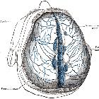
Normal
intracranial calcifications • Falx cerebri calcification - Ganzer Fall bei Radiopaedia

Normal
intracranial calcifications • Tentorium cerebelli calcifications - Ganzer Fall bei Radiopaedia

Basal ganglia
calcification • Basal ganglia calcification - senile - Ganzer Fall bei Radiopaedia

Normal
intracranial calcifications • Bochdalek's flower basket - Ganzer Fall bei Radiopaedia

Normal
intracranial calcifications • Orbital roof fracture and intracerebral hemorrhage - Ganzer Fall bei Radiopaedia

Normal
intracranial calcifications • Normal CT head - Ganzer Fall bei Radiopaedia
Normal intracranial calcifications can be defined as all age-related physiologic and neurodegenerative calcifications that are unaccompanied by any evidence of disease and have no demonstrable pathological cause.
The most common sites include:
- pineal gland
- seen in 2/3 of the adult population and increases with age
- calcification over 1 cm in diameter or under nine years old may be suggestive of a neoplasm
- habenula
- it has a central role in the regulation of the limbic system and is often calcified with a curvilinear pattern a few millimeters anterior to the pineal body in 15% of the adult population
- choroid plexus
- a very common finding, usually in the atrial portions of the lateral ventricles (choroid glomus)
- calcification in the third or fourth ventricle or patients less than nine years of age is uncommon
- basal ganglia
- are usually incidental idiopathic findings that have an incidence of ~1% (range 0.3-1.5%) and increases with age
- usually, demonstrate a faint punctuate or a coarse conglomerated symmetrical calcification pattern
- see basal ganglia calcification for specific differential
- falx, dura mater or tentorium cerebelli
- occur in ~10% of the elderly population
- dural and tentorial calcifications are usually seen in a laminar pattern and can occur anywhere within the cranium
- petroclinoid ligaments
- common age-related degeneration sites and usually have laminar or mildly nodular patterns
- superior sagittal sinus
- common age-related degeneration sites and usually have laminar or mildly nodular patterns
- dentate nuclei of cerebellum
- hippocampus
See also
Siehe auch:
- zerebrale Verkalkungen
- intrakranielle Verkalkungen
- Basalganglienverkalkungen
- Sinus sagittalis superior
- Glandula pinealis
- habenula
- Verkalkungen Plexus choroideus
- globus pallidus calcification
- Ligamentum petroclinoideum
- verkalkte Meningen
und weiter:


 Assoziationen und Differentialdiagnosen zu normale intrakranielle Verkalkungen: