pericallosal lipoma


 nicht verwechseln mit: Falxlipom
Pericallosal lipomas are fat-containing lesions occurring in the interhemispheric fissure closely related to the corpus callosum, which is often abnormal. It is the most common location for an intracranial lipoma.
Epidemiology
Pericallosal lipomas are rare, found in only 1 in 2,500 to 1 in 25,000 autopsies .
Clinical presentation
Approximately 50% of patients present with seizures . The tubulonodular variety (see below) is usually associated with more severe and extensive abnormalities and thus is more frequently symptomatic.
With the increasing use of antenatal sonography more and more cases are being detected incidentally in utero.
Pathology
The pathogenesis of a pericallosal lipoma is considered to be the result of an abnormal persistence and differentiation of the meninx primitiva into lipomatous tissue . Typically resorption occurs between the 8and the 10week of gestation.
Radiographic features
Pericallosal lipomas can be grouped into two distinct types based on imaging:
- tubulonodular
- curvilinear
Tubulonodular lipoma
Tubulonodular pericallosal lipomas are the more common variety. They are rounded or lobular and usually measure >2 cm in thickness. They are anteriorly situated and are associated with extensive callosal and often fronto-facial anomalies. The tubulonodular variety can extend into the choroid plexus / lateral ventricles .
Curvilinear lipoma
Curvilinear pericallosal lipomas are usually thin, elongated and curvilinear along the corpus callosum margin. They usually measure <1 cm in thickness and are more posteriorly situated. The corpus callosum is only mildly hypoplastic .
In a small minority of pericallosal lipomas, a connection with extracranial subcutaneous lipomas is seen. This may be through a skull defect (cranium bifidum) in which case the masses are continuous with each other, or via a thin fibrous-lipomatous stalk with an apparently intact skull vault .
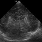
Ultrasound
Ultrasound demonstrates the characteristic appearance of fat: a hyperechoic midline mass in the region of the corpus callosum .
CT
CT is diagnostic, demonstrating fat density mass (-80 to -110HU) . Additionally, the tubulonodular variety may show peripheral curvilinear calcification sometimes referred to as the bracket sign on coronal reformatted images.
The anterior cerebral vessels can be seen coursing through or above the mass and may have associated vascular malformations or aneurysm formation . CT angiography may thus be indicated.
MRI
MRI is the modality of choice to characterize not only the extent of the lipoma but also the frequently associated agenesis/dysgenesis of the corpus callosum.
Not surprisingly these masses follow signal intensity of fat on all sequences :
- T1
- markedly hyperintense
- demonstrates signal attenuation on fat suppression sequences
- T2
- high signal, but marginally lower than CSF
- demonstrates signal attenuation on fat suppression sequences but not on FLAIR
- T1 C+ (Gd): no enhancement
- GE/EPI/SWI: the presence of peripheral calcification leads to blooming
Additionally, peripheral chemical shift artifact may be prominent in some sequences.
Again, as vascular abnormalities are associated with these lesions, careful examination of the vessels is essential (best seen on T2 FSE sequences).
Treatment and prognosis
No specific treatment is usually required , although seizures if present need medical management. In the rare cases where a cranium bifidum is present then surgical repair may be necessary.
Prognosis is good, but the degree of disability is variable, ranging from severe neurological dysfunction and debilitating seizures to completely asymptomatic.
Differential diagnosis
In general, there is little differential as few intracranial masses have significant fatty components. The differential includes:
- intracranial dermoid cyst
- intracranial teratoma
- fatty falx cerebri: especially to be considered in curvilinear type
- rare lipomatous transformation of neoplasm: PNET, ependymoma, glioma
See also
Siehe auch:
- Falxlipom
- Ependymom
- intrakranielle Lipome
- intrakranielles Fett
- intrakraniale Teratome
- Tumor im Corpus callosum
- intrakranielle Dermoidzyste
- classification system for midline abnormalities of the brain
- Primitiver neuroektodermaler Tumor
- ribbon lipoma with corpus callosal dysgenesis
- Pai-Syndrom (Lippenspalte - faziale Polypen - ZNS-Lipome)
und weiter:


 Assoziationen und Differentialdiagnosen zu Balkenlipom:


