persistent falcine sinus


The falcine sinus is a normal fetal structure located within the falx cerebri that drains the deep cerebral venous system to the superior sagittal sinus. Normally, it involutes after birth.
Gross anatomy
The falcine sinus drains into the superior sagittal sinus and usually arises from the vein of Galen or the anterior part of the straight sinus, coursing through the posterior third of the falx cerebri . The name has also described an anomalous venous structure in the anterior falx that drained the inferior sagittal sinus to the superior sagittal sinus .
Variant anatomy
Falcine sinus is present after birth in 2-5% of individuals . In this situation, it may be referred to as persistent falcine sinus or recanalized falcine sinus, depending on the presumed etiology.
Persistent falcine sinus in adults is usually isolated, without other intracranial anomalies . However, it is also the most common venous anomaly associated with vein of Galen aneurysmal malformation in children, particularly if the straight sinus is absent, thrombosed, or rudimentary . In this situation, the aneurysmally enlarged median prosencephalic vein (MPV) drains into the falcine sinus.
Recanalization of the falcine sinus occurs most commonly with tumors (mainly meningioma) compressing adjacent venous structures, followed by venous thrombosis (mainly superior sagittal and transverse sinuses) .
Radiographic features
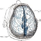
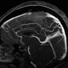
Venography (by CT, MR, or catheter-based angiography) easily demonstrates the finding on a sagittal section or lateral projection.
The caliber of falcine sinus observed after birth is highly variable, ranging 2-17 mm (mean 7 mm) . The morphology of the falcine sinus has been grouped into 3 categories, in decreasing order of frequency :
- arch-like: curved vessel protruding towards the anterior aspect
- stick-like: straight vessel
- branch-like: bifurcating vessel
Siehe auch:


 Assoziationen und Differentialdiagnosen zu persistierender Sinus falcinus: