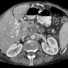
Plattenepithelkarzinom des Pankreas

Challenges in
managing upper gastrointestinal bleeding secondary to primary squamous cell carcinoma of the pancreas: a case report and literature review. Abdominal pelvic CT scan showing a pancreatic body and tail SCC with multiple liver metastases. A Axial view demonstrating multiple hypoattenuating liver metastases with the largest lesion in segment 8 measuring 68 × 64 × 63 mm (arrow). B Sagittal view demonstrating the pancreatic mass compressing on the fundus and proximal gastric body with a small gaseous locule suggesting direct gastric invasion with possible fistulous formation (arrow), C coronal view showing the pancreatic SCC invading superior mesenteric vein (SMV), splenic vein (SV) and abutting the confluence of the portal vein (PV) by more than 180° (arrows)

Challenges in
managing upper gastrointestinal bleeding secondary to primary squamous cell carcinoma of the pancreas: a case report and literature review. EUS demonstrating a large irregular hypoechoic paragastric lesion from the lesser curvature of the gastric body. EUS guided fine needle biopsy (FNB) was performed for histology

Primary
squamous cell carcinoma of the pancreas with a large pseudocyst of the pancreas as the first manifestation: a rare case report and literature review. Abdominal CT revealed pancreatitis with a pseudocyst (red arrow) with a diameter measuring 7 cm

Primary
squamous cell carcinoma of the pancreas: a case report and review of the literature. Positron emission tomography-computed tomography scan showing multiple liver metastasis immediately post-pancreaticoduodenectomy and prior to first-line palliative chemotherapy.
Plattenepithelkarzinom des Pankreas
Pankreastumoren Radiopaedia • CC-by-nc-sa 3.0 • de
There are numerous primary pancreatic neoplasms, in part due to the mixed endocrine and exocrine components.
Classification
Classification based on function
- exocrine: ~99% of all primary pancreatic neoplasms
- pancreatic ductal adenocarcinoma ~90-95%
- cystic neoplasm
- intraductal papillary mucinous neoplasm (IPMN)
- endocrine: were previously referred to as islet cell tumors because they were thought to have originated from the islets of Langerhans, however, new evidence suggests that these tumors originate from pluripotential stem cells in ductal epithelium
- non-syndromic
- syndromic
- mesenchymal tumors
- although the great majority of both benign and malignant pancreatic neoplasms arise from pancreatic epithelial cells, mesenchymal tumors, while rare, can derive from the connective, lymphatic, vascular, and neuronal tissues of the pancreas
- they account for 1-2% of all pancreatic tumors and are classified according to their histologic origin
- other
Exocrine tumors
- ductal adenocarcinoma is by far the most common primary tumor, usually of the head (65%) and has a very poor prognosis.
- cystic neoplasms are further divided into (with some overlap):
- unilocular
- intraductal papillary mucinous neoplasms (IPMN)
- serous cystadenoma uncommonly uni/macrolocular
- macrocystic multilocular
- mucinous cystic neoplasm: usually body and tail
- intraductal papillary mucinous neoplasms (IPMN)
- serous cystadenoma uncommonly uni/macrolocular
- microcystic
- serous cystadenoma: usually head. 30% have a central scar
- cystic with a solid component
- macrocystic tumors can have solid component as well
- pancreatic adenocarcinoma may undergo cystic degeneration (8%)
- unilocular
- generally solid
See also: cystic pancreatic mass: differential diagnosis
Endocrine tumors
Endocrine tumors of the pancreas are divided into:
- functional: ~85%
- insulinoma: most common, 10% are malignant
- gastrinoma: second most common, 60% malignant
- glucagonoma: 80% malignant
- VIPoma: 75% malignant
- somatostatinoma: 75% malignant
- non-functional: ~15%
- third most common
- 85-100% malignant
- usually larger, as a result of lack of hormonal activity, the clinical presentations are usually delayed till they become large
Mesenchymal tumors
Account for 1-2% of all pancreatic tumors and are classified according to their histologic origin :
These are further discussed at pancreatic mesenchymal neoplasms
Classification based on location
Head
- ductal adenocarcinoma
- intraductal papillary mucinous neoplasms (IPMN)
- serous cystadenoma
- pancreatoblastoma (rare and in children)
Body and tail
Intraductal
- intraductal papillary mucinous neoplasm (IPMN)
- pancreatic intraductal neoplasia (PanIN)
- intraductal tubular neoplasm (ITN)
- intraductal tubulopapillary neoplasm (ITPN)
- intraductal tubular adenoma (ITA)
- intraductal tubular carcinoma (ITC)
Siehe auch:

 Assoziationen und Differentialdiagnosen zu Plattenepithelkarzinom des Pankreas:


