retroperitoneales Liposarkom


Retroperitoneal liposarcoma is a subtype of liposarcoma and is a malignant tumor of mesenchymal origin that may arise in any fat-containing region of the body. It is one of the most common primary retroperitoneal neoplasms.
Epidemiology
Most cases occur in patients at the 5-7 decades of life, with no gender predilection .
Liposarcomas represent the most common variety of malignant retroperitoneal tumor.
Pathology
Histology
There are five histological types:
- well-differentiated: ~55%, low grade
- lipoma-like
- inflammatory
- sclerosing
- myxoid: ~30%, low-to-intermediate grade
- pleomorphic: high grade
- round cell: high grade
- dedifferentiated: high grade
Metastatic disease is haematogenous and the extent of metastases is related to the histological grade of the tumor.
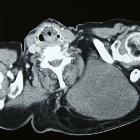
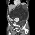
Radiographic features
CT
- varying amount of fat and soft tissue
- from purely fat with rare thin septa (usually low-grade lesions) to a very heterogeneous mass with extensive amounts of soft tissue component (usually high-grade lesions)
- multiple septa
- enhancing soft tissue components
MRI
- myxoid type:
- T2: hyperintense - myxoid gelatinous components
- C+ (Gd): delayed post-contrast enhancement
Treatment and prognosis
The primary treatment option is resection if possible. However, local recurrence is common and occurs in two-thirds of patients. This is usually a sign of incomplete resection and highlights the difficulty in discriminating liposarcomas from normal retroperitoneal fat.
Differential diagnosis
- retroperitoneal leiomyosarcoma
- retroperitoneal undifferentiated pleomorphic sarcoma (previously known as malignant fibrous histiocytoma MFH)
- retroperitoneal fibrosarcoma
- retroperitoneal lipoma
- extremely rare; therefore, should not be entertained in the differential for retroperitoneal fat-containing lesions
- exophytic renal angiomyolipoma (AML)
- presence of a large vessel extending into the renal cortex suggestive of AML; liposarcomas are hypovascular
- claw sign
- renal parenchymal defect at the site of tumor contact strongly favors the diagnosis of exophytic angiomyolipoma
- calcifications suggest liposarcoma
Siehe auch:
- Myelolipom Nebenniere
- Angiomyolipom der Niere
- Liposarkom
- retroperitoneale Tumoren
- retroperitoneales Leiomyosarkom
- retroperitoneale Sarkome
- pleomorphes undifferenziertes Sarkom des Retroperitoneums
und weiter:


 Assoziationen und Differentialdiagnosen zu retroperitoneales Liposarkom: