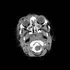
Subperiosteal abscess of the orbit

Toddler with
a runny nose and right orbital swelling. Axial CT with contrast of the orbits (above) shows opacification of the ethmoid sinuses and sphenoid sinus along with right preseptal soft tissue swelling. There is a fluid collection with rim enhancment medially next to the wall of the right ethmoid sinus which is better seen on the coronal CT (below). The diagnosis was sinusitis with preseptal cellulitis and a subperiosteal abscess.

School ager
with left eye swelling Axial CT with contrast of the orbits shows opacification of the left ethmoid sinus and bilateral sphenoid sinuses along with pre and post-septal inflammation of the left orbit. A lenticular fluid collection with rim enhancement is present along the medial wall of the left orbit.The diagnosis was orbital cellulitis with a subperiosteal abscess.
Subperiosteal abscess of the orbit occurs as a complication of acute sinusitis.
Clinical presentation
Patients can present with pain, visual disturbance, proptosis and/or chemosis.
Pathology
Bacteria can extend via neurovascular foramina or bony dehiscences. More commonly occurs from ethmoidal sinusitis, extending into the orbit via the lamina papyracea but can also occur secondary to frontal sinusitis.
Radiographic features
CT
- low density collection in a juxta-osseous location adjacent to a sinus with features of sinusitis
- may not enhance peripherally
Complications
- orbital cellulitis
- arterial or venous thrombosis
Siehe auch:
und weiter:

 Assoziationen und Differentialdiagnosen zu subperiostaler Abszess der Orbita:


