thorakale Sarkoidose


Sarkoidose
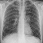
der Lunge im Stadium der Fibrose (Stadium 4 nach Scadding, Stadium 3 nach Wurm) im Röntgenbild des Thorax.

Sarcoidosis
(thoracic manifestations) • Sarcoidosis - Ganzer Fall bei Radiopaedia

Pulmonary
sarcoidosis. Multiple enlarged homogeneous mildly enhancing Mediastinal lymph nodes are seen in left paratracheal (green arrows) region.

Pulmonary
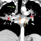
sarcoidosis. Conglomerate bilateral perihilar masses are seen extending along peribronchovascular interstitium radiating to periphery (star). Note enlarged subcarinal (orange arrow) and bilateral hilar regions (yellow arrows).
Pulmonary
sarcoidosis. Conglomerate bilateral perihilar masses are seen extending along peribronchovascular interstitium radiating to periphery (star). Note enlarged right paratracheal (green arrows), subcarinal (orange arrow) and bilateral hilar regions (yellow arrows)


Pulmonale
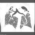
Sarkoidose im Röntgenbild und der Computertomographie coronar rekonstruiert.

Pulmonary
sarcoidosis. Sarcoid cluster consists of a cluster of multiple micronodules seen in the peripheral lung (blue arrow).

Pulmonary
sarcoidosis. Multiple well-defined, small, rounded nodules are seen predominantly in the upper and middle lobes. The nodules are seen in subpleural location (blue arrows) and peribronchovascular interstitum (green arrows) suggestive of perilymphatic distribution.

Pulmonary
sarcoidosis. Multiple well defined, small, rounded nodules are seen predominantly in the upper lobes. The nodules are seen along the fissures (yellow arrow), in subpleural location (blue arrow) suggestive of perilymphatic distribution.

Pulmonary
sarcoidosis. Multiple well-defined, small, rounded nodules are seen predominantly in the upper lobes. The nodules are seen along the fissures (yellow arrow), in subpleural location (blue arrow) suggestive of perilymphatic distribution.

Pulmonary
sarcoidosis. Conglomerate perihilar masses (star) extending along the peribronchovascular interstitium. Note the bronchi traversing through these masses without their obliteration. Mulitple well-defined nodules are seen along the peribronchovascular interstitium (green arrows) and subpleural areas (blue arrow).

Pulmonary
sarcoidosis. Multiple enlarged homogeneous mildly enhancing mediastinal lymph nodes are seen in subcarinal (orange arrow) and bilateral hilar regions (yellow arrows). Note right perihilar mass radiating to periphery (star).

Sarcoidosis
(thoracic manifestations) • Sarcoidosis (gross pathology) - Ganzer Fall bei Radiopaedia
Sarcoidosis
(thoracic manifestations) • Sarcoidosis - Ganzer Fall bei Radiopaedia

Pulmonary
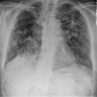
sarcoidosis. Reticulo-nodular pattern is seen in right perihilar region and left mid zone (blue arrows). Few nodular opacities are also seen in left upper zone and adjacent to left costo-phrenic angle (circle).

Sarkoidose in
der Computertomographie: Querschnitt durch den Thorax im Bereich der Aufzweigungen der Bronchien (der Hili) mit vielen vergrößerten Lymphknoten (Pfeile).
Sarkoidose
der Lunge im Stadium der Fibrose (Stadium 4 nach Scadding, Stadium 3 nach Wurm) im Röntgenbild des Thorax.


Nodular lung
lesions are reported in up to 4% of cases of sarcoidosis. The main differential diagnosis is usually metastatic neoplasm.

An AP chest
xray showing the typical nodularity of sarcoidosis in a person with sarcoidosis

Sarkoidose
bei einem 36jährigen Mann. Befall von Leber und Milz sowie hier der Lunge. Die Computertomographie axial zeigt multiple solide Herde, die primär an ein Malignom denken ließen, zumal die Befunde in der PET-CT stark stoffwechselaktiv waren.


Pulmonary
sarcoidosis. Multiple enlarged homogeneous mildly enhancing mediastinal lymph nodes are seen in left paratracheal (green arrows) and prevascular regions (blue arrows).

Sarcoidosis
(thoracic manifestations) • Sarcoidosis - Ganzer Fall bei Radiopaedia

Sarcoidosis
(thoracic manifestations) • Sarcoidosis - Ganzer Fall bei Radiopaedia

Sarcoidosis
(thoracic manifestations) • Sarcoidosis - Ganzer Fall bei Radiopaedia

Sarcoidosis
(thoracic manifestations) • Sarcoidosis - Ganzer Fall bei Radiopaedia

Sarcoidosis
(thoracic manifestations) • Sarcoidosis - stage 1 - Ganzer Fall bei Radiopaedia

Sarcoidosis
(thoracic manifestations) • Pulmonary sarcoidosis - Ganzer Fall bei Radiopaedia
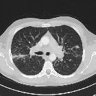
Sarcoidosis
(thoracic manifestations) • Sarcoidosis - Ganzer Fall bei Radiopaedia

Sarcoidosis
(thoracic manifestations) • Pulmonary sarcoidosis - Ganzer Fall bei Radiopaedia

Sarcoidosis
(thoracic manifestations) • Pulmonary sarcoidosis - Ganzer Fall bei Radiopaedia

Sarcoidosis
(thoracic manifestations) • Sarcoidosis - Ganzer Fall bei Radiopaedia

Sarcoidosis
(thoracic manifestations) • Medastinal sarcoidosis - Ganzer Fall bei Radiopaedia

Sarcoidosis
(thoracic manifestations) • Pulmonary sarcoidosis - stage IV - Ganzer Fall bei Radiopaedia

Sarcoidosis
(thoracic manifestations) • Sarcoidosis - Ganzer Fall bei Radiopaedia

Sarcoidosis
(thoracic manifestations) • Sarcoidosis - perilymphatic nodular pattern - Ganzer Fall bei Radiopaedia

Sarcoidosis
(thoracic manifestations) • Sarcoidosis (alveolar type) - Ganzer Fall bei Radiopaedia

Sarcoidosis
(thoracic manifestations) • Sarcoidosis - ground glass opacity and perilymphatic nodular pattern - Ganzer Fall bei Radiopaedia

Sarcoidosis
(thoracic manifestations) • Pulmonary sarcoidosis with fibrosis - Ganzer Fall bei Radiopaedia

Sarcoidosis
(thoracic manifestations) • Pulmonary sarcoidosis - Ganzer Fall bei Radiopaedia

Sarcoidosis
(thoracic manifestations) • Sarcoidosis - stage II - Ganzer Fall bei Radiopaedia

Sarcoidosis
(thoracic manifestations) • Sarcoidosis - Ganzer Fall bei Radiopaedia

Sarcoidosis
(thoracic manifestations) • Sarcoidosis - Ganzer Fall bei Radiopaedia

Sarcoidosis
(thoracic manifestations) • Sarcoidosis - Ganzer Fall bei Radiopaedia

Sarcoidosis
(thoracic manifestations) • Pulmonary sarcoidosis - Ganzer Fall bei Radiopaedia

Sarcoidosis
(thoracic manifestations) • Sarcoidosis - Ganzer Fall bei Radiopaedia

Sarcoidosis
(thoracic manifestations) • Sarcoidosis - stage II - Ganzer Fall bei Radiopaedia

Sarcoidosis
(thoracic manifestations) • Sarcoidosis - stage I - Ganzer Fall bei Radiopaedia

Sarcoidosis
(thoracic manifestations) • Pulmonary sarcoidosis - Ganzer Fall bei Radiopaedia

Sarcoidosis
(thoracic manifestations) • Sarcoidosis - fibrocystic changes - Ganzer Fall bei Radiopaedia

Sarcoidosis
(thoracic manifestations) • Sarcoidosis - stage I - Ganzer Fall bei Radiopaedia

Sarcoidosis
(thoracic manifestations) • Thoracic sarcoidosis - Ganzer Fall bei Radiopaedia
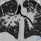
Pulmonary
sarcoidosis. Mulitple well-defined nodules are seen along the peribronchovascular interstitium (green arrows) and subpleural areas (blue arrow).
thorakale Sarkoidose
Siehe auch:
- Bronchopneumogramm
- Tuberkulose
- Lymphangiosis carcinomatosa
- Sarkoidose
- Lungensarkoidose
- Neurosarkoidose
- Lungenfibrose
- Honigwabenmuster
- Aspergillom
- pulmonale Hypertonie
- miliare Lungenherde
- gewöhnliche interstitielle Pneumonie (UIP)
- Sarkoidose Stadien
- Traktionsbronchiektasen
- zentrilobuläre Lungennoduli
- Eierschalenverkalkungen
- musculoskeletal manifestations of sarcoidosis
- löfgren syndrome
- abdominelle Manifestationen der Sarkoidose
- Garland Trias
- chest radiograph classification of pulmonary sarcoidosis
- kardiale Beteiligung bei Sarkoidose
- sarcoid galaxy sign
- lambda sign
- sarcoidosis (general article)
- interlobular septae
- differential of chronic airspace opacities
- eggshell
- stage of disease
- differential of interlobular septal thickening
- secondary lobules

 Assoziationen und Differentialdiagnosen zu thorakale Sarkoidose:
gewöhnliche
interstitielle Pneumonie (UIP)


