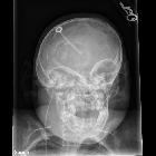
Trachealatresie

Newborn with
respiratory distress. CXR AP and lateral (above) shows a nasogastric tube with its tip in the mid-esophagus. AP image from an UGI exam performed by injecting the nasogastic tube (below left) shows simultaneous opacification of the tracheobroncial tree and esophagus. Lateral image from the UGI exam (below right) shows the atretic origin of the trachea (black arrow). The tip of the endotracheal tube (which is in the esophagus) is at approximately the same level.The diagnosis was tracheal atresia.

Failed
resuscitation of a newborn due to congenital tracheal agenesis: a case report. Floyd"s classification of tracheal agenesis.

Two case
reports of unexpected tracheal agenesis in the neonate: 3 C’s beyond algorithms for difficult airway management. Chest CT scans (sagittal (a), horizontal (b) and axial (c)) show the absence of the trachea and bronchi (B) arising from the oesophagus (OE) via a fistula (arrow ↖)

Two case
reports of unexpected tracheal agenesis in the neonate: 3 C’s beyond algorithms for difficult airway management. a Sagittal chest CT scan showing long-segment agenesis of the trachea, only oesophagus (OE) visible. b Horizontal chest CT scan displaying the fistula (arrow ↗) from the oesophagus (OE) to a ventral distal tracheal pouch (blind proximal ending) at the level of thoracic vertebrae 4–5

The role of
CT in the diagnosis of tracheal agenesis: a case report. Endotracheal tube in the right main bronchus after passing through the fistula between short distal tracheal segment and oesophagus.

The role of
CT in the diagnosis of tracheal agenesis: a case report. CT demonstrates placement of endotracheal tube in the right main bronchus after passing through the fistula between distal tracheal segment and oesophagus. Normal main bronchi and a dilated distal oesophagus(E) are demonstrated.

The role of
CT in the diagnosis of tracheal agenesis: a case report. CT demonstrates the endotracheal tube in the proximal esophagus. The trachea is absent.

The role of
CT in the diagnosis of tracheal agenesis: a case report. CT demonstrates the endotracheal tube in the proximal esophagus. The trachea is absent.

The role of
CT in the diagnosis of tracheal agenesis: a case report. Floyd classification of tracheal agenesis [2]
Tracheal atresia (TA) is an extremely rare anomaly and refers to a congenital absence of the trachea.
Epidemiology
There may be a greater male predilection .
Pathology
Tracheal atresia falls under the spectrum of laryngeal-tracheo-bronchial atresia which in turn results either from an obstructing lesion (i.e. cartilaginous bar) or vascular insult with atresia of the airways which occurs during intrauterine development.
Associations
Associated anomalies can be present in up to 90% of cases .
- congenital high airways obstruction syndrome (CHAOS): some authors consider this as synonymous with tracheal atresia although the syndrome can occur with a tracheal stenosis or laryngeal atresia/stenosis
- Fraser syndrome
- esophageal atresia with tracheo-esophageal fistula : a tracheal atresia can occur in some of the sub types
- polyhydramnios
- VACTERL association
Radiographic features
Antenatal ultrasound
- may show uniformly increased echogenic enlarged lung +/- pleural effusions +/- fetal ascites
- markedly dilated fluid-filled bronchi may be seen
- may also show ancilliary sonographic features such as the presence of polyhydramnios.
- the fetal cardio-thoracic circumference ratio is decreased due to larger lung volumes
Differential diagnosis
General differential considerations include:
- laryngeal atresia: sometimes can be impossible to differentiate on imaging
- severe congenital tracheal stenosis
Siehe auch:
- Ösophagusatresie
- Bronchialatresie
- Fraser-Syndrom
- congenital high airways obstruction syndrome
- Larynxatresie
- Floyd Klassifikation der Trachealagenesie
und weiter:

 Assoziationen und Differentialdiagnosen zu Trachealatresie:


