Varianten des hinteren Atlasbogens


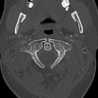
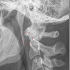
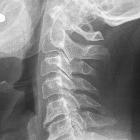
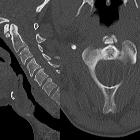
Congenital anomalies of the posterior arch of the atlas (C1) are relatively common anomalies. They may range from partial defects presenting as clefts to complete absence of the posterior arch (aplasia).
These anomalies are classified according to Currarino (see below). It should not be confused with Currarino triad (an inherited congenital disorder of the sacrum and anus or rectum).
Epidemiology
The reported incidence in one larger series from 1930 was 4% . Estimates from a recent extensive review range from 0.7-3% .
Clinical presentation
Clinical presentation is highly variable. While most are asymptomatic and present as an incidental finding on imaging studies performed after trauma or for other reasons, severe symptoms such as intermittent tetraparesis after minor cervical trauma have been described in the literature .
Not surprisingly, higher morphological groups (especially C and D, owing to a posterior tubercle) tend to be symptomatic, either post-trauma or intermittent to chronic. For example, symptoms due to impingement of the cord by a posterior tubercle during extension is well described in the literature . However, post-traumatic symptoms have been described in the milder forms .
Classification
Congenital anomalies of the posterior atlas arch can be classified according to a system proposed by Currarino et al. in 1994 consisting of a combination of morphology and clinical presentation . At the time of writing (August 2016) the Currarino classification remains the most widely used:
- morphological types
- type A: failure of posterior midline fusion of the two hemiarches
- type B: unilateral defect
- type C: bilateral defects
- type D: absence of the posterior arch, with persistent posterior tubercle
- type E: absence of the entire arch, including the tubercle
- clinical subgroups
- 1: incidental imaging finding, asymptomatic
- 2: neck pain or stiffness after trauma to the head or neck
- 3: chronic symptoms referable to the neck
- 4: various chronic neurological problems
- 5: acute neurological symptoms following minor cervical trauma
Type A and subgroup 1 are by far the commonest (approximating 80% of cases) and are encountered in 4% of the general population . In contrast, all other morphological types (B to E) are encountered in only 0.69% of the population .
Pathology
Embryology
This rare anomaly is a developmental failure of chondrogenesis (lack of chondrification). In the embryological period C1 is usually formed from three primary ossification centers:
- an anterior center developing into the anterior tubercle
- two lateral centers giving rise to the lateral masses and posterior arch
In ~2% of the population, an additional ossification center develops in the posterior midline, subsequently forming into a posterior tubercle.
During ossification different anomalies can develop, comprising:
- median cleft(s) of the posterior arch
- varying degrees of posterior arch dysplasia
- either with or without the presence of posterior tubercle (see above)
Fusion of ossicles usually occurs during age 3 to 5 years. Incomplete posterior fusion may even be normal in children up to 10 years old .
Associations
Association with congenital anomaly of the posterior arch of the atlas has been reported in several disorders, including:
Mechanism
One theory hypothesizes that a reduced distance between occiput and spinous processes in extension causes spinal cord compression by inward buckling of the ligaments and that this is a possible mechanism of acute or chronic injury.
Another hypothesis for neurological signs is secondary hypertrophy of the ligaments and dura due to a partial or complete absence of the posterior arch.
Treatment and prognosis
Anomalies of the posterior arch of C1 are usually considered benign , but may give rise to severe neurological compromise. Especially groups C and D (i.e. isolated posterior ossicle) may be considered a risk factor for neurological morbidity rather than a developmental variant of normal .
In cases of doubt (e.g. post-traumatic symptoms not clearly related to the anomaly, type C or D without symptoms) imaging studies may include :
- cervical lateral plain radiography, optimally as flexion and extension study
- cervical CT with multiplanar reconstruction and/or 3D
- cervical MRI, possibly with the neck in extension
- including medullary and soft-tissue sequences to depict spinal cord changes and mapping of ligamentous structures
For asymptomatic cases, no treatment or follow-up is needed, as considered a benign anatomical variant. Some authors including Currarino suggest advising patients with type C and D to avoid contact sports. Prevention of cumulative damage to the cord by surgery at an early stage may also be prudent in these types with a posterior tubercle. In cases with symptomatic compression, surgery with excision of the posterior arch is considered curative .
Differential diagnosis
When patients with arch anomalies present with trauma, the radiographs may be confusing and misleading. Hence thorough knowledge of these abnormalities is essential to avoid misinterpretation as an osteolytic lesion, fracture or dislocation, e.g. atlantoaxial subluxation.
See also
Siehe auch:
- Ossikel am vorderen Atlasbogen
- Atlasbogenspalt
- Foramen arcuale
- Varianten des vorderen Atlasbogens
- Spalt am hinteren Atlasbogen
- Laminektomie Atlas
- Currarino's classification of anomalies of posterior arch of atlas
- aplasia of the posterior arch of the atlas vertebra
und weiter:

 Assoziationen und Differentialdiagnosen zu Varianten des hinteren Atlasbogens:


