congenital uterine anomalies

Uterus
bicornis bicollis mit gemeinsamer Portioöffnung: Das Cavum uteri ist vollständig getrennt, es gibt aber noch gemeinsames Myometrium (sonst Uterus didelphys); Septum in nahezu der kompletten Zervix. MRT T2, links 2 Schichten paracoronar, rechts 3 Schichten axial in verschiedenen Höhen.

Müllerian
duct anomalies • Arcuate uterus - Ganzer Fall bei Radiopaedia

Female
teenager with right lower quadrant pain. Sequential coronal T2 MRI without contrast images of the pelvis show the uterus to consist of a single fluid filled dilated right horn (above) which empties into the vagina (below).The diagnosis was Mullerian duct anomaly in the form of right unicornuate uterus.

Müllerian
duct anomalies • Mayer-Rokitansky-Küster-Hauser syndrome - Ganzer Fall bei Radiopaedia

Mature cystic
ovarian teratoma • Septate uterus with ovarian pathologies - Ganzer Fall bei Radiopaedia

Uterus •
Uterine anatomical abnormalities (illustrations) - Ganzer Fall bei Radiopaedia

The clinical
relevance of anatomical variants!. The distal third of the two hemivaginas (arrows) separated by a band of fibrous tissue (arrowhead); the obstructed right hemivagina is already dilated, containing heterogeneous debris, which indicates distal obstruction.

Accessory and
cavitated uterine mass (ACUM)- A rare Mullerian anomaly. STIR axial image showing thick-walled lesion contiguous with the left lateral uterine wall.

Accessory and
cavitated uterine mass (ACUM)- A rare Mullerian anomaly. T1 fat suppressed axial image shows bright signal in the central part of the lesion consistent with signal intensity of blood degradation products.

Müllerian
duct anomalies • Müllerian duct development - Gray's anatomy illustration - Ganzer Fall bei Radiopaedia

Septate
uterus • Uterine anatomical abnormalities (illustrations) - Ganzer Fall bei Radiopaedia

Müllerian
duct anomalies • Bicornuate uterus with solitary kidney - Ganzer Fall bei Radiopaedia

Uterine
duplication anomalies • Bicornuate uterus - Ganzer Fall bei Radiopaedia

Uterine
duplication anomalies • Septate uterus with ovarian pathologies - Ganzer Fall bei Radiopaedia

Renal
agenesis • Unilateral renal agenesis and class II Mullerian duct anomaly - Ganzer Fall bei Radiopaedia

Uterine
duplication anomalies • Bicornuate uterus with triamniotic dichorionic triplet pregnancy (3D ultrasound) - Ganzer Fall bei Radiopaedia

Müllerian
duct anomalies • Unicornuate uterus with renal anomaly - Ganzer Fall bei Radiopaedia

Uterus
subseptus in der Computertomographie links axial und rechts in der Ebene des Uterus rekonstruiert. Zusätzlich zystischer Tumor mit abgebildet.

Müllerian
duct anomalies • Unicornuate uterus - type B - Ganzer Fall bei Radiopaedia

Septate
uterus • Septate uterus - Ganzer Fall bei Radiopaedia

Unicornuate
uterus • Unicornuate uterus (3D ultrasound) - Ganzer Fall bei Radiopaedia

The clinical
relevance of anatomical variants!. Sagittal transabdominal US image shows an elliptical echogenic mass, which represents the vaginal cavity markedly distended with blood products (haematocolpos). The uterus extends from the superior aspect of the mass.

The clinical
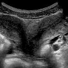
relevance of anatomical variants!. Transverse transabdominal US image shows uterus didelphys, with two uterine horns and two separated endometrial cavities.

The clinical
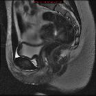
relevance of anatomical variants!. MR image shows a dilated hemivagina whose content has signal characteristics of blood, forming a blind-ending pouch secondary to obstruction.

The clinical
relevance of anatomical variants!. A large haematocolpos centrally (arrow), a finding that corresponds to the obstructed right hemivagina. Mild dilatation of the right endometrial cavity and a nondistended left endometrial cavity are also seen.

The clinical
relevance of anatomical variants!. Coronal T2-weighted image of a uterus didelphys, obtained in plane with the uterus, shows two widely divergent uterine horns (arrows) separated by a deep fundal cleft.

The clinical
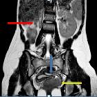
relevance of anatomical variants!. Coronal fast spin-echo T2-WI shows a solitary left kidney, confirming renal agenesis with bowel loops in the right renal fossa, which is ipsilateral to the obstructed hemivagina.

The clinical
relevance of anatomical variants!. Transverse oblique T2-WI obtained with fat suppression demonstrate communication of the right uterine horn (curved arrow) with the dilated left hemivagina (arrow); the appearance of the left uterine horn is normal.

Female
teenager with abdominal pain. Sequential axial CT with contrast of the abdomen shows a fluid-filled bladder in the anterior aspect of the pelvis that has a soft tissue structure posterior to it, believed to be the uterus, that is composed of what appear to be left and right horns.The diagnosis was Mullerian duct anomaly in the form of a uterus didelphys or a bicornuate uterus.
There are many classification systems for congenital utero-vaginal anomalies. These include:
- Buttram and Gibbons classification
- American Fertility Society (AFS) classification
- Modified Rock and Adam - AFS classification
Modified Rock and Adam - AFS classification
This classification divides congenital uterine anomalies into four main types:
- class I: dysgenesis of Müllerian ducts
- includes agenesis or hypoplasia of the müllerian duct derivatives: the uterus and upper two-thirds of the vagina
- the most common form is the Mayer-Rokitansky-Kuster-Hauser syndrome which is the combined agenesis of the uterus, cervix, and upper portion of the vagina
- class II: disorders of vertical fusion
- these anomalies are due to failure of fusion of the müllerian system with the sinovaginal bulb
- they include cervical dysgenesis and obstructive and nonobstructive transverse vaginal septa
- class III: disorders of lateral fusion
- describes anomalies that result in a duplicated or partially duplicated reproductive tract
- these disorders are due to impaired fusion and/or septal resorption of fusing Müllerian ducts attempting to form the uterus, cervix, and upper vagina
- it includes anomalies due to failure of fusion of the paired müllerian ducts (as in didelphic and bicornuate uteri) and failure of midline septum resorption after fusion (as in septate uterus)
- disorders due to lateral fusion defects are further subclassified into
- a: symmetric non-obstructive forms seen in five types: unicornuate, bicornuate, didelphic, septate, and DES-related uteri
- b: asymmetric obstructive forms seen in three types: unicornuate uterus with obstructed horn, double uterus with unilaterally obstructed horn, and double uterus with unilaterally obstructed vagina
- class IV: unusual configurations and combinations of defects
Siehe auch:
- Uterus
- Uterus didelphys
- Müllerian duct anomaly classification
- Uterus arcuatus
- Uterus septus
- Uterus bicornis
- Mayer-Rokitansky-Küster-Hauser syndrome
- Uterus unicornis
- Müller-Gang
- transverses Vaginalseptum
- obstruierte Hemivagina und ipsilaterale renale Anomalie (OHVIRA) Syndrom
- Vaginalseptum
- Duplikatur des Uterus
- Uterusagenesie
- Müller-Gang-Persistenzsyndrom
- class IV Mullerian duct anomaly
und weiter:
- Hysterosalpingographie
- Vagina
- cervical incompetence
- velamentous cord insertion
- MRKHS
- fetal akinesia / hypokinesia sequence
- Entwicklungsanomalien der Plazenta
- Müller Gang Embryologie
- infertility in the exam
- gynäkologisch radiologisches Curriculum
- haematometrocolpos
- MRKH
- Anomalien des urogenitalen Systems
- longitudinales Vaginalseptum
- primary amenhorrea due to Müllerian duct anomalies
- mullerian duct cyst in an infant
- hydrometrocolpos in a neonate
- accessory and cavitated uterine mass
- Uterus subseptus
- vaginal duplication
- persistent Müllerian duct syndrome

 Assoziationen und Differentialdiagnosen zu Fehlbildungen der Gebärmutter:
obstruierte
Hemivagina und ipsilaterale renale Anomalie (OHVIRA) Syndrom


