bronchopleurale Fistel

Teenager with
a bacterial pneumonia not responsive to oral antibioticsAxial CT without contrast of the chest shows consolidation of the right lower lobe with multiple air pockets within it giving the appearance of necrotic lung, a large loculated right empyema that contains an air pocket, and one of the air pockets in the right lower lobe appears to be in continuity with the air pocket in the pleural space suggesting the presence of a bronchopleural fistula.The diagnosis was bacterial pneumonia with a bronchopleural fistula.

Teenager with
a complicated bacterial pneumoniaCXR AP shows a large right pleural effusion with an air-fluid level within it – a hydropneumothorax – that is being drained by a chest tube.The diagnosis was right-sided bronchopleural fistula.

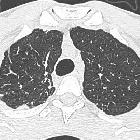
Toddler with
respiratory distress. CXR AP (above) shows diffuse bilateral airspace disease and a right sided pneumothorax that is almost completely drained by a right chest tube. Axial CT with contrast of the chest (below left) shows air and fluid and a chest tube in the right pleural space and an enhancing rim sign of the pleura. There is also a bronchopleural fistula from the superior segment of the right lower lobe to the necrotic lung and pleural space (below right).The diagnosis was bronchopleural fistula and pleural empyema in a patient with streptococcus pneumonia.

Bronchopleural
fistula • Bronchopleural fistula - Ganzer Fall bei Radiopaedia

Bronchopleural
fistula • Bronchopleural fistula - Ganzer Fall bei Radiopaedia

Bronchopleural
fistula • Post-operative left bronchopleural fistula - Ganzer Fall bei Radiopaedia

Bronchopleural
fistula • Bronchopleural fistula - postoperative complication - Ganzer Fall bei Radiopaedia

Bronchopleural
fistula • Bronchopleural fistula secondary to tuberculosis - Ganzer Fall bei Radiopaedia

Bronchopleural
fistula • Clagett thoracotomy with bronchopleural fistula - Ganzer Fall bei Radiopaedia
Bronchopleural fistulas are communications between the bronchial tree and the pleural space.
Pathology
They are usually divided as:
- central: when the fistula involves the trachea or a lobar bronchus
- peripheral: when a distal airway, either segmental bronchi or the lung parenchyma, communicates to the pleural space
Etiology
- postoperative complication of pulmonary resection: considered by far the most common cause, with a reported incidence from 1.5 to 28% after pulmonary resection
- may rarely be caused by pleuroparenchymal fibroelastosis
- lung necrosis complicating infection or infarction
- traumatic
- pneumatoceles
- iatrogenic (eg. thoracic tube insertion, lung biopsy, toracocentesis, and nasogastric tube malpositioning)
- lung neoplasms
- tumor extension into the pleural space
- tumor necrosis after chemotherapy or radiotherapy
Radiographic features
Plain radiograph
On chest radiography, the features that may be seen include:
- steady increase in intrapleural airspace
- appearance of a new intrapleural gas-pleural fluid collection - i.e. a hydropneumothorax. The gas-fluid level typically extends to the chest wall and shows unequal linear dimensions on orthogonal views conforming to the pleural space
- changes in an already present gas-fluid level
- development of tension pneumothorax
- a drop in the gas-fluid level exceeding 2 cm (if the patient has no chest tube in place)
CT
CT is considered the imaging technique of choice for visualizing and characterizing bronchopleural fistulae . CT may show:
- pneumothorax
- hydropneumothorax
- pneumomediastinum
- underlying lung pathology
- demonstration of an actual fistulous communication
Nuclear medicine
Radioaerosol scanning (e.g. xenon ventilation nuclear scintigraphy) has been successfully used in the evaluation of bronchopleural fistulas. A variety of radioactive tracers may be used, including:
- technetium-99m (99mTc) albumin colloid fog inhalation
- 99mTc sulfur colloid
- 99mTc-labeled diethylenetriamine pentaacetate, krypton, and xenon
- single photon emission tomography using radiolabeled aerosol inhalation. If there is fistula the radioactive tracer will equilibrate between the postpneumonectomy or pleural space and the airways after inhalation
Siehe auch:
- Seropneumothorax
- Pneumatozele
- Pleuraempyem
- pulmonale Tuberkulose
- Lungeninfarkt
- pleuroparenchymale Fibroelastose
- Lungennekrose
und weiter:


 Assoziationen und Differentialdiagnosen zu bronchopleurale Fistel: