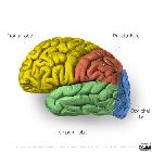
Frontallappen


The frontal lobe is by far the largest of the four lobes of the cerebrum (other lobes: parietal lobe, temporal lobe, and occipital lobe), and is responsible for many of the functions which produce voluntary and purposeful action.
Gross anatomy
The frontal lobe is the largest lobe accounting for 41% of the total neocortical volume . The frontal lobe resides largely in the anterior cranial fossa, lying on the orbital plate of the frontal bone. Its most anterior part is known as the frontal pole and extends posteriorly to the central (Rolandic) sulcus which separates it from the parietal lobe. Posteroinferiorly it is separated from the temporal lobe by the lateral sulcus (Sylvian fissure), although not seen from the surface is the insular cortex which is hidden deep to the lateral sulcus . The interhemispheric fissure separates its medial surface from the contralateral frontal lobe.
The frontal lobe is roughly pyramidal in shape, with three cortical surfaces:
Lateral surface
The lateral surface is curved, conforming to the inner surface of the frontal and parietal bones. It is divided into four gyri, which in reality are more 'regions' than true gyri, in that each is convoluted and divided by smaller incomplete sulci, which in turn are separated from each other by three main sulci .
- gyri
- superior frontal gyrus
- middle frontal gyrus
- inferior frontal gyrus
- precentral gyrus
- sulci
- superior frontal sulcus
- inferior frontal sulcus
- precentral sulcus
Although each gyrus and sulcus are discussed individually, a brief overview is presented here.
- most medial and superior part of the frontal lobe
- extends from the frontal pole anteriorly to the precentral sulcus posteriorly (separating it from the precentral gyrus)
- laterally it is separated from the middle frontal gyrus by the superior frontal sulcus
- runs parallel to the superior frontal gyrus, from frontal pole to precentral sulcus
- superomedially it is separated from the superior frontal gyrus by the superior frontal sulcus
- inferolaterally it is separated from the inferior frontal gyrus by the inferior frontal sulcus
- runs parallel to the middle frontal gyrus, from the lateral border of the orbital gyri anteroinferiorly
- superiorly it is separated from the middle frontal gyrus by the inferior frontal sulcus
- a large part of the inferior frontal gyrus forms the frontal operculum, which covers the insular cortex
- contains Broca's area
- runs in a roughly coronal plane, angling anteriorly as it passes from vertex down towards the lateral sulcus
- anteriorly it is separated from the posterior parts of the superior, middle and inferior frontal gyri by the precentral sulcus
- posteriorly it is separated from the parietal lobe by the central sulcus
- contains the primary motor cortex
Medial surface
The medial surface of the frontal lobe, abutting the falx in the midline, is primarily divided by the curving cingulate sulcus, which parallels the outer outline of the corpus callosum.
Above the cingulate sulcus is the medial continuation of the superior frontal gyrus, which is usually divided into two parts by a short ascending branch from the cingulate sulcus.
- medial frontal gyrus
- anterior to the ascending branch of the cingulate sulcus
- paracentral lobule
- posterior to the paracentral sulcus (the ascending branch of the cingulate sulcus)
- anterior to the marginal branch of the cingulate sulcus
- NB: the most posterior part of the paracentral lobule includes the medial continuation of the postcentral gyrus, and thus is nominally part of the parietal lobe.
Below the cingulate sulcus is the cingulate gyrus, which is variably included partially as part of the frontal lobe, or sometimes considered part of the limbic lobe .
The frontal pole wraps from the lateral surface and onto the medial surface. The inferior part of the medial surface of the frontal lobe is composed of two relatively sizable gyri which run horizontally from the frontal pole towards the lamina terminalis, separated from the later by a small relatively complicated region known as the septal area .
- gyrus rectus: forms the inferior most part of the lobe, wrapping around onto the inferior surface (see below)
- rostral gyrus: runs immediately above gyrus rectus, separated from it by the inferior rostral sulcus
- septal area: located below the rostrum of the corpus callosum and posteroinferior most extent of the cingulate gyrus, anterior to the lamina terminalis, and posterior to the rostral gyrus and gyrus rectus
Inferior surface
The inferior surface of the frontal lobe is the smallest cortical surface of the lobe, located anterior to the stem of the Sylvian fissure, lying on the floor of the anterior cranial fossa. It is divided into two parts, by two sulci.
Most medial, and running anteroposteriorly is the olfactory sulcus, which separates the long thin medial gyrus rectus (straight gyrus), from the larger orbital gyri .
The orbital gyri are in turn divided by the H-shaped orbital sulcus, into four gyri, two located above and below the transverse part of the "H" (the anterior and posterior orbital gyri), and two located on either side of the "H" (the medial and lateral orbital gyri) .
Relations
- anterior: frontal bone
- superiorly: frontal bone (anteriorly), coronal suture, and parietal bone (posteriorly)
- posterior: central sulcus and parietal lobe
- inferolaterally: lateral sulcus and temporal lobes
- inferior: floor of anterior cranial fossa
Arterial supply
- middle cerebral artery (MCA): lateral frontal lobe
- anterior cerebral artery (ACA): medial frontal lobe
Neurological deficits
The following neurological deficits occur with unilateral or bilateral lesions of the frontal lobes :
- deficits arising from unilateral dominant side lesions:
- Broca aphasia: expressive aphasia
- problems with repetition: lesions affecting the arcuate fasciculus
- destruction of frontal eye field: impaired gaze to the contralateral side
- deficits arising from unilateral non-dominant side lesions:
- hemiparesis/hemiplegia
- deficits arising from bilateral lesions:
- intellectual impairment
- personality change
- disinhibition
- apathy
- abulia (loss of drive)
- urinary incontinence
- Foster Kennedy syndrome (lesions in the olfactory groove region): anosmia, ipsilateral optic atrophy, and contralateral papilledema
Siehe auch:

 Assoziationen und Differentialdiagnosen zu Frontallappen:


