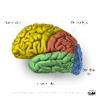
Temporallappen


The temporal lobe is one of the four lobes of the brain (along with the frontal lobe, parietal lobe, and occipital lobe), and largely occupies the middle cranial fossa.
Gross anatomy
The temporal lobe is the second largest lobe, after the larger frontal lobe, accounting 22% of the total neocortical volume . The lobe extends superiorly to the Sylvian fissure, and posteriorly to an imaginary line; the lateral parietotemporal line, which separates the temporal lobe from the inferior parietal lobule of the parietal lobe superiorly and the occipital lobe inferiorly. The middle cranial fossa forms its anterior and inferior boundaries.
The temporal lobe can be divided into two main sections:
- neocortex (sometimes referred to simply as temporal lobe)
- lateral and inferolateral surfaces
- comprised of standard cerebral cortex
- mesial temporal lobe (sometimes referred to as the limbic lobe)
- including the hippocampus, amygdala, parahippocampal gyrus (see: mesial temporal lobe)
Sulci and gyri
The temporal lobe is divided into five gyri by four sulci which are oriented largely parallel to the Sylvian fissure. Unfortunately, not all gyri and sulci extend the full length of the lobe and as such not all are present at each angled coronal section. Furthermore, nomenclature is variable.
The order from superolateral to inferomedial is:
Blood supply
Arterial supply
The temporal lobe receives blood from both the internal carotid artery and the vertebrobasilar artery :
- internal carotid system
- anterior choroidal artery
- supplies the anterior segment of parahippocampal gyrus, the uncus and the amygdala
- middle cerebral artery
- supplies superior and inferior temporal gyri and temporal pole
- several temporal branches arise from the MCA although there is considerable variation in anatomical arrangement
- temporopolar artery
- anterior temporal artery
- middle temporal artery
- posterior temporal artery
- anterior choroidal artery
- vertebrobasilar system
- supplies the inferior surface of the temporal lobe via the temper-occipital artery
Venous drainage
Venous drainage occurs via two routes :
- blood from the neocortex travels anteriorly to the superficial middle cerebral vein, then into the inferior anastomotic vein (vein of Labbé) which joins the transverse sinus
- blood from the mesial temporal lobe goes to the posterior choroidal vein which goes on to form the internal cerebral vein, eventually becoming the great cerebral vein (vein of Galen), continuing into the straight sinus
Neurological deficits
The following neurological deficits occur with unilateral or bilateral lesions of the temporal lobes :
- deficits arising from unilateral lesions involving the dominant hemisphere:
- alexia: acquired dyslexia (inability to read)
- agraphia: inability to write
- acalculia: inability to calculate
- Wernicke's dysphasia: receptive dysphasia
- nominal dysphasia: inability to name objects (lesions involving the posterior-superior temporal lobe)
- contralateral homonymous superior quadrantanopia: 'pie in the sky' visual field defect (due to disruption of Meyer's loop which dips into the temporal lobe)
- deficits arising from unilateral lesions involving the non-dominant hemisphere:
- contralateral homonymous superior quadrantanopia
- prosopagnosia: failure to recognize faces
- irritative lesions involving either lobe can give rise to the following:
- formed visual hallucinations
- complex-partial seizures
- memory disturbances (e.g. déjà vu and other memory disturbances)
Related pathology
Siehe auch:
- Hippocampus
- mesial temporal lobe
- lateral parietotemporal line
- Temporallappenepilepsie
- Frontallappen
- Hirnlappen
- Okzipitallappen
- Tumoren des Temporallappens
und weiter:

 Assoziationen und Differentialdiagnosen zu Temporallappen:


