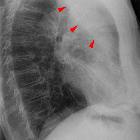
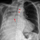
lung collapse


Lung atelectasis (plural: atelectases) refers to collapse or incomplete expansion of pulmonary parenchyma.
Terminology
Atelectasis may be used synonymously with collapse, but some authors reserve the term "atelectasis" for partial collapse, not inclusive of total atelectasis of the affected part of lung or of whole lung collapse.
Classification
Atelectasis is a radiopathological sign which can be classified in many ways. The aim of each classification approach is to help identify possible underlying causes together with other accompanying radiological and clinical findings.
Atelectasis can be subcategorised based on underlying mechanism, as follows:
- resorptive (obstructive) atelectasis
- occurs as a result of complete obstruction of an airway
- no new air can enter the portion of the lung distal to the obstruction and any air that is already there is eventually absorbed into the pulmonary capillary system, leaving a collapsed section of the affected lung
- because the visceral and parietal pleura do not separate in resorptive atelectasis, traction is created, and if the loss of volume is considerable, mobile thoracic structures may be pulled toward the side of volume loss ("mediastinal shift")
- potential causes of resorptive atelectasis include obstructing neoplasms, mucus plugging in asthmatics or critically ill patients and foreign body aspiration
- resorptive atelectasis of an entire lung ("collapsed lung") can result from complete obstruction of the right or left main bronchus
- passive (relaxation) atelectasis
- occurs when contact between the parietal and visceral pleura is disrupted
- the three most common specific etiologies of passive atelectasis are pleural effusion, pneumothorax and diaphragmatic abnormality
- compressive atelectasis
- occurs as a result of any thoracic space-occupying lesion compressing the lung and forcing air out of the alveoli
- cicatrisation atelectasis
- occurs as a result of scarring or fibrosis that reduces lung expansion
- common etiologies include granulomatous disease, necrotizing pneumonia and radiation fibrosis
- adhesive atelectasis
- occurs from surfactant deficiency
- depending on etiology, this deficiency may either be diffuse throughout the lungs or localized
- gravity dependent atelectasis (dependent atelectasis)
- in the most dependent portions of the lungs due to the weight of the lungs
- osteophyte-induced adjacent pulmonary atelectasis and fibrosis
Atelectasis can also be subcategorised by morphology:
- linear (a.k.a. plate, band, discoid) atelectasis: a minimal degree of collapse as seen in patients who are not taking deep breaths ("splinting"), such as postoperative patients or patients with rib fracture or pleuritic chest pain; this is very common
- round atelectasis: classically associated with asbestos exposure
or by anatomical extent:
- lung atelectasis: complete collapse of one lung
- lobar atelectasis: collapse of one or more lobes of a lung.
- segmental atelectasis: collapse of one or more individual pulmonary segments
Radiographic features
Vary depending on the underlying mechanism and type of atelectasis
Plain radiograph / CT
Direct signs of atelectasis
- displacement of interlobar fissures
- crowding together of pulmonary vessels
- crowded air bronchograms (does not apply to all types of atelectasis; can be seen in subsegmental atelectasis due to small peripheral bronchi obstruction, usually by secretions; if the cause of the atelectasis is central bronchial obstruction, there will usually be no air bronchograms)
Indirect signs of atelectasis
- pulmonary opacification
- shifting granuloma (or any other previously documented lesion, used as a reference for comparison)
- compensatory hyperexpansion of the surrounding or contralateral lung
- displacement of the heart, mediastinum, trachea, hilum
- elevation of the diaphragm
- propinquity of the ribs
Resorptive (obstructive) atelectasis
- increased density (opacity) of the atelectatic portion of lung
- displacement of the fissures toward the area of atelectasis
- upward displacement of hemidiaphragm ipsilateral to the side of atelectasis
- crowding of pulmonary vessels and bronchi in region of atelectasis
- +/- compensatory overinflation of unaffected lung
- +/- displacement of thoracic structures (if atelectasis is substantial)
Linear (plate, discoid, subsegmental) atelectasis
- relatively thin, linear densities in the lung bases oriented parallel to the diaphragm (known as Fleischner lines)
Ultrasound
The sonographic morphology of atelectatic lung may resemble hepatic parenchyma, often referred to as "tissue-like" or "hepatized" in appearance. Distinguishing features of atelectasis by etiology may appear as follows:
- compressive atelectasis is most often visualized in the costophrenic recess bordered by a disproportionately large pleural effusion
- low-level, homogenous echogenicity with few to no air bronchograms
- margins are usually regular with a triangular shape
- a shred sign may be present at the transition to aerated lung
- obstructive atelectasis
- early static air bronchograms due to distal air trapping
- as the air is resorbed, bronchi may fill with fluid resulting in anechoic, tubular structures known as fluid bronchograms
- maybe differentiated from blood vessels with color flow Doppler
History and etymology
Atelectasis comes from the Greek words 'ateles' and 'ektasis' translating to 'incomplete stretching or expansion'.
Siehe auch:
- Mittellappenatelektase
- Mediastinalshift
- Unterlappenatelektase
- Rundatelektase
- Oberlappenatelektase
- lobar collapse
- Surfactant-Mangelsyndrom
- segmental atelectasis
- Kompressionsatelektase
- Plattenatelektase
und weiter:
- Bronchopneumogramm
- right middle lobe consolidation
- left lower lobe collapse
- right upper lobe collapse
- Pseudopneumoperitoneum
- left upper lobe collapse
- right lower lobe consolidation
- pulmonary opacification
- Unterlappenatelektase rechts
- Parenchymband
- subpleural line
- tracheobronchiale Papillomatose
- juvenile laryngotracheal papillomatosis
- small round cell lung carcinoma with neuroendocrine features
- Mediastinalverbreiterung
- verkalkte Atelektasen
- Veil-like opacity

 Assoziationen und Differentialdiagnosen zu Atelektase:


