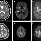
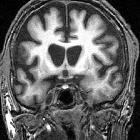
T2 hyperintense basal ganglia (mnemonic)

Syndrome of
uremic encephalopathy and bilateral basal ganglia lesions in non-diabetic hemodialysis patient: a case report. Brain MRI performed upon admission. T2WI showed symmetrical hyperintense non-hemorrhagic lesions in the entire bilateral basal ganglia (arrows), and corona radiata lesions showing mild diffusion restriction

Magnetic
resonance imaging of arterial stroke mimics: a pictorial review. A 42-year-old woman with hypoglycaemia presenting with disturbance of consciousness after abdominal surgery. DWI shows hyperintensities in the basal ganglia and the splenium of the corpus callosum (a, c, arrows) with no restriction (b) and no arterial occlusion (d)

Lentiform
fork sign in a uremic patient with a high anion gap metabolic acidosis with seizures: a case report from North West of Ireland. Showing CT Scan and MRI Brain images. a CT-brain showing bilateral symmetrical hypodensity in the basal ganglia. b MRI Brain axial FLAIR sequence. c MRI brain coronal T2 sequences both showing bilateral symmetrical swollen lentiform nuclei with a hyperintense T2/FLAIR signal rim delineating the boundaries of the putamen

Syndrome of
uremic encephalopathy and bilateral basal ganglia lesions in non-diabetic hemodialysis patient: a case report. Brain MRI performed upon admission. T2FLAIR showed symmetrical hyperintense non-hemorrhagic lesions in the entire bilateral basal ganglia (arrows) and corona radiata lesions showing mild diffusion restriction

Basal ganglia
T2 hyperintensity • Wilson disease - Ganzer Fall bei Radiopaedia

Basal ganglia
T2 hyperintensity • Osmotic demyelination syndrome - Ganzer Fall bei Radiopaedia

Basal ganglia
T2 hyperintensity • CNS lymphoma - Ganzer Fall bei Radiopaedia

Basal ganglia
T2 hyperintensity • Huntington disease - Ganzer Fall bei Radiopaedia

Basal ganglia
T2 hyperintensity • Basal ganglia chronic hypoxic ischemic disease - Ganzer Fall bei Radiopaedia

Magnetic
resonance imaging patterns of paediatric brain infections: a pictorial review based on the Western Australian experience. T2 hyperintensity in basal ganglia and/or thalami (Pattern 5). Case 1: 15-year-old child with headache, nausea and vomiting. CSF was positive for cryptococcus. T2-weighted imaging (a) demonstrated bubble-like lesions within the basal ganglia (relating to gelatinous pseudocysts), with a relatively smaller component of restricted diffusion (b). Case 2: 14-year-old child with malaise for 2 weeks. CSF was positive for cryptococcus. T2-weighted imaging (c) demonstrated bubble-like lesions within the basal ganglia and thalami, with a relatively smaller component of restricted diffusion (d)
A helpful mnemonic to recall the causes of T2 hyperintense basal ganglia is:
- LINT
Mnemonic
- L: lymphoma
- I: ischemia
- N: neurodegenerative conditions
- T: toxins
See also
For a more detailed differential please see T2 hyperintense basal ganglia article.
Siehe auch:
- Lymphom
- Morbus Wilson
- Leigh-Syndrom
- ADC abnormality of the basal ganglia
- Creutzfeldt-Jakob-Krankheit
- Kohlenmonoxidintoxikation
- Chorea Huntington
- Morbus Behçet ZNS-Manifestationen
- decreased T1 signal in the basal ganglia
- decreased T2 signal in the basal ganglia
- Methanolintoxikation
- basal ganglia signal abnormalities
- Lentiform fork sign (basal ganglia)
- Methylmalonazidurie
- increased T1 signal in the basal ganglia
und weiter:
- Basalganglienverkalkungen
- Globus pallidus
- Stammganglieninfarkt
- Putamen
- basal ganglia T1 hyperintensity
- Basalganglien
- Einblutung in die Basalganglien
- basal ganglia T2 hypointensity
- eye of tiger sign
- Nucleus caudatus
- Japanische Enzephalitis
- hyperdense Basalganglien
- respiratory chain metabolic toxins
- Enzephalitis durch Flaviviren
- signal abnormalities in the basal ganglia
- T2 hyperintense Ponsläsionen
- ethylene glycol toxicity
- signalintensity MRI basalganglia
- T2 hyperintenses Putamen
- T2 Stammganglien

 Assoziationen und Differentialdiagnosen zu T2 hyperintense Basalganglien:
ADC
abnormality of the basal ganglia


