antenatal features of Down syndrome
Antenatal screening of Down syndrome (and other less common aneuploidies) should be available as a routine component of antenatal care. It allows families to either adjust to the idea of having a child with the condition or to consider termination of pregnancy.
For a general description of Down syndrome and its postnatal manifestations, please refer to the article on Down syndrome.
Maternal age
There is a strong association between the incidence of Down syndrome and maternal age. Background risk based on maternal age is incorporated into both the serum screening based risk calculations and into the calculation of increased risk in the presence of a soft marker in 2 trimester (see below).
Maternal serum screening
Please refer to the antenatal screening for a general discussion of the avenues of screening and diagnosis in the antenatal period.
1 trimester
Combined serum screening has an approximately 85% detection rate, with 5% false positive rate .
Serological markers: MSS1
- maternal beta hCG
- higher than chromosomally normal fetuses
- the difference increases with gestation
- PAPP-A
- lower than chromosomally normal fetuses
- difference decreases with gestation: therefore not commonly used as a second-trimester test
2 trimester
A triple screening/quadruple screening is done in high-risk cases. However combined screening test is most preferred. Detection rate for trisomy 21 is at approximately 80% with a false positive rate of ~5% .
Serological markers: MSS2
- maternal free beta-hCG: higher than chromosomally normal fetuses
- inhibin A: higher than chromosomally normal fetuses
- AFP: lower than chromosomally normal fetuses
- unconjugated estriol (uE3): lower than chromosomally normal fetuses
Ultrasound
First trimester
Nuchal translucency: thickness depends on the size of the fetus (CRL), but in general it is considered abnormal if >3 mm.
There is evidence that the inclusion of nasal bone measurement improves the specificity of 1 trimester data .
Ductus venosus flow is another parameter that has been reported to further increase the sensitivity of 1 trimester screening .
Second trimester
Approximately 30% of babies with Down syndrome have detectable abnormalities on the mid-trimester ultrasound .
Soft markers
Soft markers are sonographic findings that do not in themselves cause any adverse outcomes. However, they are seen more frequently in fetuses with an abnormality. This article addresses the soft markers that are specific to Down syndrome. For a general discussion, please refer to the article on soft markers.
The markers are as follows :
- echogenic intracardiac focus
- positive LR: 5.83
- negative LR: 0.8
- isolated LR: 0.95
- ventriculomegaly
- positive LR: 27.5
- negative LR: 0.94
- isolated LR: 3.8
- nuchal fold thickness >6 mm
- positive LR: 23.3
- negative LR: 0.8
- isolated LR: 3.79
- echogenic bowel
- positive LR: 11.44
- negative LR: 0.9
- isolated LR: 1.65
- hypoplastic/absent nasal bone
- positive LR: 23.3
- negative LR: 0.46
- isolated LR: 6.58
- shortened humerus
- positive LR: 4.8
- negative LR: 0.7
- isolated LR: 0.78
- mild pyelectasis
- positive LR: 7.6
- negative LR: 0.9
- isolated LR: 1.08
- shortened femur
- positive LR: 3.72
- negative LR: 0.8
- isolated LR: 0.61
- aberrant right subclavian artery (ARSA)
- positive LR: 21.48
- negative LR: 0.7
- isolated LR: 3.9
In the presence of soft markers, the risk of Down syndrome is recalculated as new risk = baseline risk x likelihood ratio (LR). The new LR is calculated by multiplying all positive LRs (of markers present) and all negative LRs (of markers absent).
Note: if a single marker is present, then isolated LR is considered.
Structural abnormalities
The following may be present in association with Down syndrome:
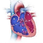
- cardiac defects
- abdominal
- duodenal atresia
- esophageal atresia
- omphalocele (more common with trisomy 18 )
- central nervous system
- craniofacial/calvarial
- other
Adjunct features
Other features that may be present, but are neither a structural abnormality or a validated soft marker
- hypoplastic 5 digit
- wide iliac angle
- shortened frontothalamic distance
- short fetal ear length
- brachydactyly
Some fetuses can develop transient abnormal myelopoiesis (TAM) particularly towards the 3trimester and can then develop fetal hepatomegaly .
Siehe auch:
- Down-Syndrom
- Brachydaktylie
- Atriumseptumdefekt
- Hydrops fetalis
- Ösophagusatresie
- nuchal translucency
- Fallot'sche Tetralogie
- fetal ventriculomegaly
- hypoplastic nasal bone
- Duodenalatresie
- abnormal ductus venosus waveforms
- Golfballphänomen
- zystisches Lymphangiom
- Omphalozele
- singuläre Nabelschnurarterie (sNSA)
- atrioventricular septal defect (AVSD)
- antenatal screening
- Ventrikelseptumdefekt
- Brachyzephalie
- shortened femur
- short maxilla
- transient abnormal myelopoiesis (TAM)
- echogenic bowel
- nuchal fold thickness
- shortened humerus
- mild pyelectasis
- absence of a nasal bone

 Assoziationen und Differentialdiagnosen zu antenatal features of Down syndrome:


