angiomyolipoma


Angiomyolipome
an beiden Nieren (Pfeile) in der Computertomographie. Die Tumore sind wegen ihres Fettgehaltes hypodens.

Computertomographie:
Multiple Angiomyolipome der Nieren bei Patientin mit CT-Bild einer Lymphangioleiomyomatose der Lunge: Dringender Verdacht auf Tuberöse Sklerose.

Computertomographie:
Multiple Angiomyolipome der Nieren bei Patientin mit CT-Bild einer Lymphangioleiomyomatose der Lunge: Dringender Verdacht auf Tuberöse Sklerose.

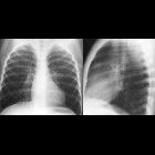
Computertomographie:
Multiple Angiomyolipomen der Nieren bei Patientin mit CT-Bild einer Lymphangioleiomyomatose der Lunge: Dringender Verdacht auf Tuberöse Sklerose.

Computertomographie:
Multiple Angiomyolipomen der Nieren bei Patientin mit CT-Bild einer Lymphangioleiomyomatose der Lunge: Dringender Verdacht auf Tuberöse Sklerose.

Renal
angiomyolipoma: Hypointens (fat isodens) lesion of right kidney in CT.

Renal
angiomyolipoma: Hypointens (fat isodens) lesion of right kidney in CT.

Teenager with
an abdominal mass. Axial CT without contrast of the abdomen shows the right kidney to be normal in size and to contain multiple small low density lesions that when measured demonstrate negative Hounsfield units. The left kidney, which is massively enlarged, also contains multiple small low density lesions as well as one extremely large low density lesion in its anterior aspect.The diagnosis was angiomyolipoma of the kidneys in a patient with tuberous sclerosis.

Angiomyolipoma
• Exophytic renal angiomyolipoma - Ganzer Fall bei Radiopaedia

Teenager with
tuberous sclerosis and abdominal pain. Axial CT with contrast of the abdomen shows a small round low density lesion in the posterolateral aspect of the left kidney (above) and a larger round low density lesion in the posterior aspect of the right kidney (below). There is also an intermediate density round lesion in the anteromedial aspect of the left kidney (above).The diagnosis was angiomyolipomas of the kidneys in a patient with tuberous sclerosis with the left anteromedial lesion having microscopic fat and the left posterolateral and right posterior lesions having macroscopic fat.
Angiomyolipomas (AMLs) refer to hamartomatous lesions composed of abnormal, thick-walled vessels (i.e. angio) and varying amounts of smooth muscle–like cells (i.e. myo) and adipose tissue (i.e. lipoma) They predominantly occur in the kidney (renal angiomyolipoma) but occasionally occur in other organs such as the liver (hepatic angiomyolipoma).
Very rarely they can occur in unusual locations such as
- skin - cutaneous angiomyolipoma
- pancreas - angiomyolipoma of the pancreas
- retroperitoneum - retroperitoneal angiomyolipoma
Siehe auch:
- Myelolipom Nebenniere
- Nierenzellkarzinom
- Angiomyolipom der Niere
- Lymphangioleiomyomatose
- retroperitoneales Liposarkom
- Nephroblastom
- Angiomyolipom der Leber
- Onkozytom
- Angiomyolipom des Pankreas
und weiter:
- Lipom
- genitourinary curriculum
- Liposarkom
- junktionaler Parenchymdefekt der Niere
- xanthogranulomatöse Pyelonephritis
- Nierentumor
- Angiomyolipome der Niere bei tuberöser Sklerose
- Adenomyolipom
- bilateral giant renal angiomyolipomas
- perivascular epithelioid cell tumours
- renal cell carcinoma and multiple angiomyolipomas in a patient with tuberous sclerosis
- eingeblutetes Angiomyolipom
- retroperitoneales Hamartom
- perivaskulärer epitheloidzelliger Tumor (PECom)
- Angiomyolipom der Milz
- lipomatöse Läsionen


 Assoziationen und Differentialdiagnosen zu Angiomyolipom: