bony metastases


Skeletal metastases (a.k.a. bone metastases) are common and result in significant morbidity in patients with metastatic disease. Although the diagnosis is often straightforward, especially as in many cases there is a well-documented history of metastatic malignancy, sometimes they may mimic benign disease or other primary malignancies.
Terminology
It is important to realize that although we commonly talk about bone metastases, strictly-speaking most of these lesions are bone marrow metastases, and metastases originating in the cortical bone itself are much less common . Indeed most of the metastases we see in the cortical bone have directly invaded from the cancellous bone/bone marrow and on x-ray and CT are often not visible until cortical bone involvement has occurred .
Epidemiology
Skeletal metastases account for 70% of all malignant bone tumors, and are seen in a vast number of primary cancers, although lung cancer, breast cancer, renal cell carcinoma and prostate cancer account for approximately 80% of all skeletal metastases . This is due to not only the propensity of these tumors to metastasize to bone, but also the fact that these are some of the most common tumors.
Clinical presentation
The majority of metastases to bone are asymptomatic. Symptoms can arise in a number of scenarios :
- direct compression of adjacent structures by extraosseous soft tissue mass (e.g. cord compression)
- palpable mass
- deformity
In most cases the diagnosis of metastatic disease is already known. If no known primary exists, or there is uncertainty regarding the diagnosis (e.g. no known metastases; unusual imaging appearances) then a bone biopsy can usually allow definitive diagnosis.
Laboratory investigations are of limited value, but will often demonstrate increased serum calcium and alkaline phosphatase . Increase in hydroxyproline excretion may also be present .
Pathology
The major route of spread of tumor to bone is haematogenous, although lymphatic spread is also seen (e.g. pelvic tumors spreading to para-aortic nodes, and then directly into bone cf. the more common haematogenous spread from the same tumors) . Although direct extension of tumors in bone is also not infrequently seen (e.g. oral cavity tumors into mandible or Pancoast tumors into first rib or upper thoracic vertebrae) this is not usually what is considered metastatic disease .
Regardless of the route of spread, metastases lead to both bone loss and bone formation, in varying amounts. The former is most likely due to direct enzymatic destruction and osteoclast activation. The latter can be due to stromal bone formation (formation of bone within tumor substrate; the case in prostate cancer metastases) or reactive new bone formation which represents the normal adjacent bone's response to the presence of tumor and is similar to callus formation .
Distribution
The distribution of skeletal metastases roughly mirrors the distribution of red marrow, presumably reflecting increased blood flow in red marrow compared to yellow marrow. Thus, metastases are usually found in:
- vertebrae
- especially the posterior vertebral body, extending into the pedicles (see: vertebral metastases)
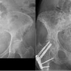
- pelvis
- proximal femur
- proximal humerus
- skull
Metastases distal to the elbow and knee are distinctly uncommon (see distal appendicular skeletal metastases).
Radiographic features
Skeletal metastases invariably incite a mixture of bone resorption and bone formation and can thus take on one of three patterns, depending on the dominant process:
Additionally, metastases can have different morphological characteristics:
- diffuse
- focal
- expansile (see: blow-out bone metastases)
Plain radiograph
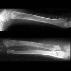
As is the case with other other bone lesions, skeletal metastases can be difficult to identify on plain films since extensive (30-50%) bone mineral loss is required before the density loss is radiographically-visible .
In many other cases the lesion is visible due to destruction of cortex, or the presence of visible sclerosis.
It is important to note that unlike primary bone tumors, in general metastases incite no or only limited periosteal reaction. The occasional exception to this general rule includes prostate cancer, some gastrointestinal malignancies, retinoblastoma and neuroblastoma .
CT
CT does not have a role in primary assessment for the presence of metastases (except for difficult areas to image such as the spine) but is excellent at defining the extent of bony involvement and in helping assess the risk of pathological fracture.
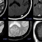
MRI
Whole body MRI is not widely used, but is highly sensitive to replacement of normal bone marrow .
Nuclear medicine
Bone scintigraphy
Bone scans are the most sensitive routine imaging modality to try and identify both sclerotic and lytic lesions . In most cases they demonstrate increased uptake (hot spot) although occasionally (in very aggressive purely lytic lesions) a photopenic defect (cold spot) may be visible. A superscan is also a possible pattern where extensive diffuse metastatic disease results in a uniform increase in uptake .
Treatment and prognosis
In general treatment can be thought of as systemic (e.g. chemotherapy or hormonal therapy) or local (e.g. radiotherapy or surgery). Pain management is also often an important part of managing patients with skeletal metastases.
A rule of thumb is that there is a high-risk of pathological fracture if the lesion is painful, >2.5 cm in size and involves >50% of the bone. There are more formal classification systems, although increasing cortical involvement is probably the most important factor :
No single statement can be made with regards to the prognosis of patients with skeletal metastases as this will vary greatly depending on the primary tumor.
Differential diagnosis
There are, unfortunately, no specific features of metastases, although often the diagnosis is straightforward in the setting of known advanced malignancy and multiple lesions.
When no history of malignancy is present, but lytic lesions are multiple in an elderly patient, the main differential is multiple myeloma.
When no helpful features are present (in other words a solitary lesion in an otherwise supposedly well patient) one needs to consider numerous entities:
- benign and malignant tumor
- infection
- trauma
- osteonecrosis
The differential can be narrowed according to specific appearances and locations:
- lytic bone metastases
- sclerotic bone metastases
- mixed lytic and sclerotic bone metastases
- expansile bony lesion
- solitary lesions
- solitary sclerotic bone lesion
- solitary lucent bone lesion: FOG MACHINES is a good place to start
- solitary lucent skull lesion


 Assoziationen und Differentialdiagnosen zu Knochenmetastasen: