Epidermoidzyste der Milz


Splenic epithelial cysts, also referred as splenic epidermoid cysts or primary splenic cysts, are unilocular fluid lesions with thin and smooth walls and no enhancement. They represent ~20% of cysts found in the spleen, and are usually an innocuous incidental imaging finding.
Note that most (~80%) simple-appearing cystic splenic lesions represent secondary cysts or pseudocysts (see Differential diagnosis section below).
Terminology
In practice, both primary and secondary cysts are often described simply as "splenic cyst" for the sake of simplicity, as often the specific etiology is not evident.
Epidemiology
They are thought to account for 20% of benign non-parasitic cysts of the spleen . There may be an increased female predilection.
Clinical presentation
The clinical presentation can vary ranging from being incidentally discovered on routine imaging to having nausea, vomiting, vague abdominal pain or rarely painful splenomegaly, particularly when the cysts are large.
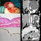
Pathology
They are congenital in origin. As “true” cysts, they have an epithelial lining formed secondary to the unfolding of peritoneal mesothelium or collections of peritoneal mesothelial cells trapped within the splenic sulci. A genetic defect of mesothelial migration is considered the cause. While most are sporadic, rarely there is a familial occurrence. They typically grow slowly and are usually large (around 10 cm) at the time of discovery. They are solitary in 80%. Peripheral calcifications are uncommon (10-15%).
Radiographic features
Ultrasound
Usually shows an anechoic to hypoechoic well defined intrasplenic lesion. Internal echoes may be present due to debris. Their margin may be echogenic with distal shadowing due to calcifications .
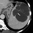
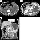
CT
Typically shows a hypoattenuating relatively well-defined intrasplenic lesion. The wall is thin and has a sharp demarcation to the splenic parenchyma. There is no rim or internal enhancement. Wall calcification may be present.
MRI
Well-defined cystic lesions with internal homogeneous fluid signal intensity:
- T1: low signal intensity
- T2: very high signal intensity
Treatment and prognosis
Small and asymptomatic cysts do not require treatment or followup. Symptomatic cysts are managed surgically.
Complications
Complications are rare and include hemorrhage, rupture and infection .
Differential diagnosis
A number of splenic lesions may appear cystic, depending on the modality:
- secondary cysts (~80% of all splenic cysts)
- splenic pseudocysts
- post-traumatic pseudocyst: the end-stage of splenic hematoma with resultant liquefactive necrosis and cystic changes
- post-infarction pseudocyst: also resultant from liquefactive necrosis
- intrasplenic pancreatic pseudocyst
- infection
- splenic pseudocysts
- other congenital cystic lesions (tend to be unilocular in a majority of cases)
- splenic bacterial abscess
- cystic splenic metastases
- splenic lymphoma: although may appear nearly anechoic, hypoechoic lymphoma tends to have less distinct margins than a cyst
- splenic peliosis
Siehe auch:


 Assoziationen und Differentialdiagnosen zu Splenic epidermoid cyst: