Granulomatosis with polyangiitis (thoracic manifestations)


Thoracic manifestations of granulomatosis with polyangiitis (GPA), which is a type of pulmonary angiitis and granulomatosis, are common; with lung involvement seen in about 95% of cases.
For a general discussion of the condition, please refer to the main article on granulomatosis with polyangiitis (GPA). For other organ-specific radiographic features, please refer to individual articles:
- granulomatosis with polyangiitis: renal manifestations
- granulomatosis with polyangiitis: upper respiratory tract manifestations
- granulomatosis with polyangiitis: CNS manifestations
- granulomatosis with polyangiitis: orbital manifestations
Granulomatosis with polyangiitis (Wegener granulomatosis), is a multi-system systemic necrotizing non-caseating granulomatous vasculitis affecting small to medium-sized arteries, capillaries and veins.
Clinical presentation
Occasionally hemoptysis due to pulmonary hemorrhage can be the presenting symptom . Cough is also reported.
Radiographic features
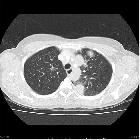
Unfortunately, the radiographic appearances of GPA are widely variable, making a diagnosis by imaging alone often difficult. Four patterns are recognized, although the first two are the most common :
- most common radiological presentation
- multiple nodules of variable size randomly distributed throughout the lungs
- bronchovascular bundles or subpleural distribution are common
- nodules may demonstrate cavitation
Pleural effusions are seen in 10-25% of cases, usually as a result of cardiac or renal involvement.
Tracheal and upper respiratory tract thickening may also be seen (see upper respiratory tract manifestations of GPA).
Plain radiograph
- chest radiographs may show multiple nodules or masses that can be extremely variable in size (from a few millimeters to many centimeters)
- although cavitation is present in ~ 50% of cases, is seen less frequently on plain film
- airspace opacities may represent consolidation or pulmonary hemorrhage
CT
Described CT features include:
- nodules or masses: variable size but typically ~2-4 cm
- multiple in 75%
- no zonal predilection
- irregularly margined
- cavitation with irregular / thick-walled cavity margins seen in ~40-50% of cases
- cavitation may be seen in 25% of nodules >2 cm
- can have a peribronchovascular or subpleural distribution
- waxing and waning even without treatment
- micronodules: centrilobular tree in bud nodules are usually related to bronchiolar wall involvement or retained blood products in the distal airways, seen in about 10% of cases
- air space consolidation
- peripheral wedge-shaped opacities (due to pulmonary infarcts)
- focal
- peribronchial
- diffuse interstitial/alveolar opacities are a more common manifestation in children
- mild bronchiectasis
- ground glass changes
- frequently as a consequence of hemorrhages
- may relate to nodules or regions of consolidation
- may be the main abnormality
- focal atelectasis from airway stenoses
- tracheobronchial wall thickening
- circumferential, can be smooth or nodular
- posterior wall of trachea is characteristically involved with no calcification
- pleural effusion
- non-specific acute or chronic fibrinous pleuritis may be rarely seen adjacent to nodular inflammatory lesions
- hilar and mediastinal lymphadenopathy: uncommon
Treatment and prognosis
For a general discussion of treatment and prognosis, please refer to granulomatosis with polyangiitis.
Complications
- superimposed pulmonary infection
- airway stenosis / tracheobronchial stenosis
- subglottic stenosis is the most common with an estimated occurrence of around ~20% (range 16-23%)
- pulmonary hemorrhage
Differential diagnosis
The differential depends on the dominant feature.
- differential of multiple pulmonary nodules
- differential of a single pulmonary nodule (uncommon presentation)
- differential of a cavitating lung mass
- differential of chronic air space consolidation
In cases of peripheral consolidation, appearances are very similar to pulmonary infarcts.
For tracheal thickening, relapsing polychondritis and tracheobronchopathia osteochondroplastica could be considered although these tend to spare the posterior wall and have calcifications .
Practical points
- although the most common manifestation of recurrent/relapsing disease is cavitary nodules, patients may relapse with a different pattern of involvement to their initial presentation
Siehe auch:
- Granulomatose mit Polyangiitis
- solitärer pulmonaler Rundherd
- Lungeninfarkt
- differential of a cavitating lung mass
- differential of chronic air space consolidation
und weiter:


 Assoziationen und Differentialdiagnosen zu Granulomatose mit Polyangiitis Manifestationen in der Lunge:

