haemangioma of the heart


Cardiac venous malformations (also known as cardiac hemangiomas) consists of a slow flow venous malformation and is composed of numerous non-neoplastic endothelial-lined thin-walled channels with interspersed fat and fibrous septae.
Terminology
It is important to note that according to newer nomenclature (ISSVA classification of vascular anomalies) these lesions are merely known as venous malformations. Having said that it is probably helpful in reports to include the word 'hemangioma' in brackets as this term is ubiquitous in the literature and most familiar to many clinicians.
Venous malformations are found throughout the body. This article focuses on cardiac cavernous hemangiomas. For a general discussion please refer to the general article on cavernous venous malformation.
Epidemiology
Cardiac hemangiomas are rare, benign lesions with an incidence of 1-10% among all detected benign heart tumors.
Clinical presentation
Mostly, cardiac hemangiomas are asymptomatic. However, they may cause a pericardial effusion, asymptomatic murmur, arrhythmia, hemopericardium or cardiac tamponade, complete heart block or even sudden death. In addition, these tumors have been incriminated for neurological manifestations.
Pathology
These vascular malformations are composed of numerous endothelial-lined thin-walled channels with interspersed fat and fibrous septa. Histological classification is according to the size of their vascular channels into three types:
Cavernous and capillary hemangioma are the most frequently reported forms, whereas a combination of all three features also is commonly seen, as well as fibrous and fatty components.
Location
Cardiac hemangiomas have been reported to occur in variable locations of the heart involving any cardiac layer, namely endo-, myo-, or pericardium. The left atrium has previously been suggested as the predominant location for cardiac tumors. However, recent evidence has shown no chamber predilection. Hence, the occurrence is possible anywhere from pericardium to the endocardium.
Associations
Cardiac hemangiomas can occur in the clinical setting of Kasabach-Merritt syndrome, which is characterized by multiple systemic hemangiomas associated with recurrent thrombocytopenia and consumptive coagulopathy.
Radiographic features
Ultrasound
On echocardiography, hemangiomas typically appear hyperechoic.
Angiography
Coronary arteriography demonstrates the blood supply to the tumor, which is characterized by a vascular blush, particularly in the capillary and arteriovenous types of hemangiomas, which exhibit rapid blood flow. Cavernous hemangiomas have large vascular spaces with a very slow flow and, therefore, do not typically enhance at angiography .
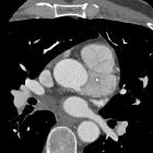
CT
Heterogeneous appearance on unenhanced chest CT, and, in most cases, intensely enhanced at CT performed after contrast material administration. Foci of calcification may also be seen be seen .
MRI
It is usually seen as a heterogeneous mass, with an intermediate-to-high signal on T1-weighted images because of slow flow. On T2-weighted images, a diffusely high signal is seen. Contrast enhancement is heterogeneous, intense and prolonged except in low-flow lesions .
Treatment and prognosis
Cardiac hemangiomas can be successfully excised, and surgical resection is the treatment of choice for symptomatic lesions or when the diagnosis is in question.The long-term outcome of patients with surgically treated symptomatic lesions is excellent. Spontaneous regression of cardiac hemangiomas have been reported, and, therefore, surgery may not always be necessary, particularly for extensive but asymptomatic hemangiomas that would require complex and potentially hazardous excision.
Complications
Potential complications include:
- development of cardiac arrhythmias
- intracavitatory growth may cause
- ventricular outflow tract obstruction
- valvular compromise
- disruption of intracardiac blood flow leading to congestive heart failure and hydrops
Differential diagnosis
Differentiation from malignancies and thrombus is very important since it greatly affects prognosis and necessity of (acute) treatment. Other differential considerations include:
- thrombus - intracardiac thrombus
- primary benign tumors
- cardiac myxoma
- cardiac fibroelastoma
- cardiac lipoma
- cardiac paraganglioma
- cardiac fibroma
- primary malignant tumors
- cardiac sarcoma(s)
- cardiac lymphoma
- cardiac malignant fibrous histiocytoma
- pericardial malignancy
- cardiac metastasis(es)
See also
Siehe auch:
- Vorhofmyxom
- intrakardiale Thromben
- papilläres Fibroelastom des Herzens
- Angiosarkom des Herzens
- kardiale Metastasen
- kardiales Fibrom
- kardiales Lipom
- primäre kardiale Tumoren
und weiter:


 Assoziationen und Differentialdiagnosen zu haemangioma of the heart:


