Hämoptyse
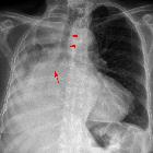
Hemoptysis (plural: hemoptyses) refers to coughing up of blood. Generally, it appears bright red in color as opposed to blood from the gastrointestinal tract which appears dark red. It is considered an alarming sign of a serious underlying etiology.
Terminology
Massive hemoptysis is referred to as expectoration of >100-600 mL of blood over a 24 hour period .
Pathology
In 90% of cases, the source of bleeding is the bronchial artery. In the remainder of cases, either the pulmonary artery or another non-bronchial artery (e.g. intercostal, internal thoracic) is the source of bleeding.
Etiology
The following are the most common causes:
- lung cancer
- bronchitis
- bronchiectasis
- tuberculosis
- aspergilloma
- pneumococcal pneumonia
- pulmonary metastasis
- pulmonary contusion
- pulmonary thromboembolism
- mitral stenosis
- iatrogenic: during catheter manipulation of pulmonary artery
Other causes:
- foreign body
- airway trauma
- lung abscess
- Goodpasture syndrome
- granulomatosis with polyangiitis
- tracheal invasion, e.g. from thyroid cancer
Rarer causes:
- arteriovenous malformation
- pulmonary endometriosis
- systemic coagulopathy
- anticoagulants
- COVID-19
Treatment and prognosis
Approach to hemoptysis
This approach can be followed for small amounts of blood or streaks of blood in sputum. The underlying cause can be life-threatening; however, it is not an emergency.
Bronchoscopy followed by a contrast-enhanced CT scan must be carried out to detect the cause. The above-mentioned common causes and certain uncommon and rare causes must be kept in mind.
Approach to massive hemoptysis
- examine the patient to rule out a non-pulmonary cause of bleeding, such as from the upper airway or gastrointestinal tract
- confirm and localize the site of bleed with bronchoscopy
- CT imaging may help in characterization of lesions if time permits
- CT arterial angiogram of the thoracic aorta should be considered in certain scenarios, particularly in patients with known cystic fibrosis
- provides definitions of the bronchial arteries anatomy and recruited aortobronchial collaterals
- CT arterial angiogram of the thoracic aorta should be considered in certain scenarios, particularly in patients with known cystic fibrosis
- DSA angiography will help localize the vessels involved and also enable embolization
- after stabilization of the patient, further imaging can be carried out and appropriate measures are taken to prevent rebleeding
Siehe auch:
und weiter:

 Assoziationen und Differentialdiagnosen zu Hämoptyse:


