hydranencephaly


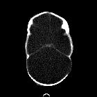
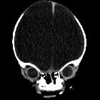
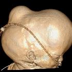
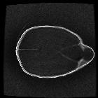

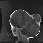
Hydranencephaly is a rare encephalopathy that occurs in-utero. It is characterized by destruction of the cerebral hemispheres which are transformed into a membranous sac containing cerebrospinal fluid and the remnants of cortex and white matter .
Porencephaly is considered a less severe degree of the same pathology .
Epidemiology
This is a rare disorder with an incidence of 0.2% in infant autopsies . It is usually sporadic. In a minority of cases, it is the consequence of the autosomal recessive Fowler syndrome.
Clinical presentation
The condition may be diagnosed prenatally using ultrasound or fetal MRI. However, it may present in neonates with seizures, respiratory failure, flaccidity or decerebrate posturing with a vegetative state .
Associations
- consequential arthrogryposis
- renal aplastic dysplasia
- polyvalvular developmental heart defects
- Fowler syndrome
- trisomy 13
- polyhydramnios
Pathology
There is complete absence of the cerebral hemispheres and often, the falx. They are replaced by a sac-like structure containing CSF surrounding the brainstem and basal ganglia .
The outer layer comprises leptomeninges while the inner layer of cortex and white matter is filled with CSF and necrotic material.
Porencephaly and hydranencephaly are considered different degrees of the same pathology. Porencephaly describes a more localized cerebral hemispheric defect, communicating with the ventricles or the cerebral surface; it tends to occur later in the developmental process .
Etiology
Five etiologies have been described:
- infarction: bilateral occlusion of the supraclinoid segment of the internal carotid arteries or of the middle cerebral arteries
- leukomalacia: an extreme form of leukomalacia formed by confluence of multiple cystic cavities
- diffuse hypoxic-ischemic brain necrosis: fetal hypoxia due to maternal exposure to carbon monoxide or butane gas may result in massive tissue necrosis with cavitation and resorption of necrotized tissue
- infection: necrotizing vasculitis or local destruction of the brain tissue secondary to intrauterine infection, e.g. congenital toxoplasmosis, cytomegalovirus, and herpes simplex (HSV) infections
- thromboplastic material from a deceased co-twin
- monochorionic twins have presented with a variety of cerebral lesions
- lesions in the recipient twin result from emboli or thromboplastic material originating from the macerated co-twin
Radiographic features
All modalities which resolve the brain parenchyma can be used to identify the features of hydranencephaly, including ultrasound (antenatal and postnatal), MRI (antenatal and postnatal), and CT. MRI is the gold standard.
In all cases, the anatomical features are the same, although they are demonstrated to a variable degree according to the abilities of each modality:
- essentially no remaining cortical tissue
- often islands of residual tissue preserved at occipital poles and orbitofrontal regions
- medial temporal tissue may be identified, as the medial temporal lobes are supplied by the basilar circulation
- preserved thalami and posterior fossa
- falx is usually present
- hemicranium is filled with fluid, in which choroid can often be identified
- antenatal ultrasound or vascular imaging demonstrate absence of middle cerebral arteries
Treatment and prognosis
Hydranencephaly is not compatible with a prolonged life after birth, with the vast majority of live births dying prior to one year of age. Termination of pregnancy is usually considered justifiable due to this reason.
Differential diagnosis
General imaging differential considerations include:
- severe obstructive hydrocephalus / fetal hydrocephalus
- cortex is identifiable albeit thinned
- macrocephaly
- middle cerebral arteries preserved
- alobar holoprosencephaly +/- very large dorsal cyst of holoprosencephaly
- usually coexisting midline facial abnormalities
- no falx
- residual rind of cortical tissue often has a cup or pancake morphology, fused across the midline anteriorly
- severe open lip schizencephaly
- focal cortical defect lined by polymicrogyic cortex
Siehe auch:
- Schizenzephalie
- Verschlusshydrocephalus
- Akranie
- Anenzephalie
- alobäre Holoprosencephalie
- Mikrohydranenzephalie
- kongenitaler Hydrozephalus
- dorsal cyst of holoprosencephaly
- Acrania anencephaly sequence
und weiter:

 Assoziationen und Differentialdiagnosen zu Hydranenzephalie:


