Medical devices in the thoracic cavity

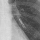
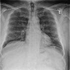
RePneu®
Coils aus Nitinoldraht zur Behandlung des Lungenemphysems im Röntgenthorax p.a.

Röntgenbild
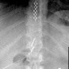
des Thorax in 2 Ebenen mit einem Carillon Mitral Contour System (Cardiac Dimensions) im Sinus coronarius. Dieses Nitinol-Device wird minimalinvasiv, katheterbasiert in den Sinus coronarius eingebracht und dort aufgespannt, um bei Mitralinsuffizienz so zu einer Annäherung der Klappenblätter zu führen und die Insuffizienz zu verringern. Zusätzlich sieht man noch einen Herzschrittmacher, eine künstliche Aortenklappe Sternalcerclagen und fraglich in der Seitprojektion einen Koronarstent.


Tiefe
Hirnstimulation bei Morbus Parkinson: Hier die Aggregate im Bereich der Brust auf einer Röntgenaufnahme des Thorax.

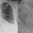
Vorhofseptumdefekt
mit Verschlusssystem: Amplatzer-Device in Seitaufnahme besser zu sehen. Zusätzlich Kardiomegalie und pulmonale Hypertonie.
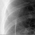
Defibrillator-Elektrode
im Röntgenbild: Die elektrische Leitung spreitzt sich in der aufgeklebten Elektrode fächerförmig auf.

Operativ
eingebrachter Spezialclip (AtriClip) auf dem linken Herzohr zur Vermeidung von Embolien aus diesem. Das Röntgenbild (Ausschnitte: links pa, rechts seitlich) zeigt zusätzlich noch eine künstliche Aortenklappe.

Z. n. TAVI
mit transapikalem Zugang. Man erkennt das Verschlusssystem an der Herzspitze (überlagert von der Sonde des ICD-Systems). Apica ASC


Medical devices in the thorax are regularly observed by radiologists when reviewing radiographs and CTs.
Extrathoracic devices
- tubing, clamps, syringes, scissors, lying on or under the patient
- rubber sheets, foam mattresses, clothing, hair braids, nipple piercings, etc. may also be visible
These devices are a common cause of artifacts and may trip the unwary, but in general are recognized for what they are.
The following are more important to be recognized by the radiologist:
- oxygen masks and ventilator support tubing
- temperature and humidity sensor attachments
- ECG electrodes/leads
- external pacemaker-defibrillator (typically seen in a cardiac patient transported by helicopter or ambulance)
- bioreactance leads (e.g. Cheetah Starling SV sensors)
- breast prostheses
- breast tissue expander (used for breast reconstruction)
- cooling blanket
- presternal peritoneal dialysis catheter
Pleural devices
- thoracostomy tubes
- usually placed anterosuperiorly to drain pneumothorax, and posteroinferiorly to drain pleural effusion
- a well-positioned tube should lie between visceral and parietal pleura, and there should not be any kinking
- to check the correct positioning, frequently AP and lateral views are required. Supplemental CT scan may also be performed.
- should not enter the interlobar fissure, else it may be blocked ; tip should not be within the lung parenchyma or subcutaneous tissue
- all drain holes should be in the pleural cavity to ensure effective drainage
- pigtail catheter: used in empyema drainage
- Heimlich valve: it is a one-way valve used for pleural space drainages, which prevents the return of gases or fluids into the pleural space
- plombage: "ping-pong ball" plombage and wax plombage (historically used for tuberculosis, but no longer)
Tracheal, bronchial and esophageal devices
- endotracheal tube
- tip of the tube should be 5 cm +/- 2 cm above the carina (carina is just caudad to the aortic arch, if not clearly visible)
- may wrongly enter right main bronchus, esophagus or even the soft tissues of the neck
- sometimes, a deliberate double-lumen ET tube is used to check differential ventilation of the two lungs
- nasogastric/nasoenteric tube/feeding tube / Dobhoff tube
- esophageal balloons (e.g. Sengstaken-Blakemore tube, Minnesota tube)
- esophageal Doppler probe
- esophageal stents
- esophageal manometer
- esophageal pH probe (seen just above gastro-esophageal junction)
- temperature probe (usually within the oropharynx or esophagus)
- tracheostomy tube
- tracheo-esophageal voice prosthesis
- bronchial stents / tracheobronchial stents (in lung transplant patients or due to obstructing tumors)
- Passy-Muir valve
- endobronchial coils
- endobronchial valves
Vascular devices
- dialysis catheters
- peripherally inserted central catheters (PICC): central portion only
- central venous catheters: central tip ideally positioned at the superior cavoatrial junction and should not enter the right atrium
- temporary non-tunnelled lines: internal jugular and subclavian lines
- tunnelled lines: e.g. Hickman line, Broviac line
- permanent, implantable access line with subcutaneous ports: e.g. Port-A-Cath, Infus-a-Port
- pulmonary artery catheter (Swan-Ganz catheter)
- left atrial catheter
- right atrial line often used postpaediatric cardiac surgery
- thoracic aortic stent
- superior vena caval stent
- superior vena caval filter
- carotid artery clamps
- cannulas of extracorporeal membrane oxygenation devices
- in the right jugular vein (in case of peripheral cannulation), rarely in the left jugular vein
- in case of central cannulation both cannulas are placed directly via central vessels into the atria
Cardiac devices
- sternal wires, plates
- cardiac prosthetic valves, C-ring annuloplasty
- cardiac conduction devices
- pacemakers (e.g. biventricular pacemaker)
- implantable cardiac defibrillators (ICD)
- coronary stents
- circulatory assist devices
- intra-aortic balloon pump (IABP)
- left ventricular assist device (LVAD) (e.g. TandemHeart percutaneous VAD)
- biventricular assist devices
- artificial heart (under development)
- temporary ventricular assist device
- atrial septal occlusion device (e.g. Amplatz closure device)
- left atrial appendage closure devices (e.g. Watchman device)
- epicardial patch
- parachute device
- cardiac restraint device
- insertable cardiac monitoring device (e.g. Reveal LINQ)
- implantable pulmonary artery pressure monitoring device (e.g. CardioMEMS)
Miscellaneous
- embolization coils
- antibiotic spacer
- pericardial drain
- insertable loop recorder
- vertebroplasty-related
- spinal rods, transpedicular screws, disc spacers, interspinous spacers
- gastric band
- reflux management system
- diaphragmatic pacemaker
- surgical clips (e.g. axillary nodal clearance)
- Nuss bar (repair of pectus excavatum)
See also
Siehe auch:
- Fremdmaterial im Röntgenbild des Thorax
- Fremdmaterial Thorax Herz
- Eventrecorder
- Herzohr Okkluder
- Mitraclips
- Herzschrittmacher
- Carillon Mitral Contour System (Cardiac Dimensions)
- Artefakte im Röntgenbild des Thorax
- Ösophagusstents
- intrakardialer Herzschrittmacher
- Plombage
- Intraaortale Ballonpumpe
- Verschlusssystem apikaler Zugang bei TAVI
- Vertebroplastie
- Kontraindikationen für eine MR-Untersuchung
- EKG-Elektroden
- Antirefluxsysteme
- abdominelle Implantate
- left ventricular assist device (LVAD)
- Defibrillator Pads aufklebbare
- EKG-Kabel
- Vagusnervstimulator
- Rückenmarkstimulation
- Koronarstent im Röntgenthorax
- Deep brain stimulators
- Defibrillator Thorax
- abdominelle Implantate und Devices
- pacemakers
und weiter:
- BH im Röntgenbild
- transcatheter aortic valve implantation (TAVI)
- Herzohr Clip
- septal occluder
- left ventricular assist device
- Okkluder
- thorakale Plombage
- Clip Thorax Herz
- MRT unmittelbar postoperativ
- biologische Aortenklappe
- Instrumentenkabel
- Artefakte im Röntgenbild
- Implantate
- RePneu Lung Volume Reduction Coil
- endoskopische Lungenvolumenreduktion
- tragbarer (wearable) Defibrillator


 Assoziationen und Differentialdiagnosen zu thorakale Implantate / Devices:


