Regenerative liver nodule


Regenerative liver nodules are a form of non-neoplastic nodules that arise in a cirrhotic liver.
Terminology
This may be slightly different from the term nodular regenerative hyperplasia, which are described histopathologically as regenerative nodules with little or no hepatic fibrosis and largely healthy hepatic architecture . When there is an accumulation of iron in the nodules, they are called siderotic nodules.
Pathology
RNs form in the setting of necrosis or regenerative nodules can be of three types :
- micronodules <3 mm
- macronodules >3 mm
- giant RNs > 5 cm (rare)
Radiographic features
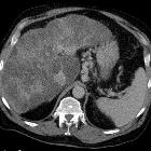
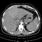
Regenerative nodules appear as round, well-defined nodules (usually in the thousands) present throughout the liver with surrounding fibrosis .
On post-contrast CT and MRI, regenerative nodules enhance similar to the normal liver parenchyma in both portal venous and hepatocellular/delayed phases, and thus may not be distinguishable in a cirrhotic liver. There is no arterial phase enhancement.
CT
Regenerative nodules are rarely visible on non-contrast CT unless they are siderotic. Siderotic regenerative nodules (containing iron) are hyperdense to liver on precontrast imaging and become isodense to liver on post contrast phases.
CT arterial portography
Contrast injection into the superior mesenteric artery (after arterial vascular access). Regenerative nodules are generally visualized as enhancing nodules surrounded by lower attenuation thin septa.
CT hepatic arteriography
Contrast injection into the common hepatic artery (after arterial vascular access). Regenerative nodules are generally visualized as nonenhancing nodules surrounded by enhancing fibrous septa.
CT hepatic arteriography is considered more sensitive than the former in depicting regenerative nodules.
MRI
RNs are common in a cirrhotic liver :
- T1: variable
- T1 in-phase/out-of-phase: loss of signal on out-of-phase if fat-containing
- T2: hypointense
- T2*: hypointense
- T1C+: usually do not enhance or enhance less than the liver parenchyma
Treatment and prognosis
Regenerative nodules may progress to dysplastic nodules or hepatocellular carcinoma .
Practical points
- fat-containing regenerative nodules are usually multiple; a solitary fat-containing nodule is concerning for dysplasia/malignancy
See also
Siehe auch:
- Leberzirrhose
- noduläre regenerative Hyperplasie
- adenomatöse Hyperplasie Leber
- dysplastische Leberknoten
und weiter:


 Assoziationen und Differentialdiagnosen zu Regenerative hepatic nodules: