Retroperitonealfibrose


 ähnliche Suchen
 siehe auch
Retroperitoneal fibrosis (RPF), is a condition that has previously been described as chronic periaortitis. It is an uncommon fibrotic reaction in the retroperitoneum that typically presents with ureteric obstruction.
The disease is part of a spectrum of entities that have a common pathogenic process consisting of an inflammatory response to advanced atherosclerosis of the abdominal aorta, combined with autoimmunologic factors:
- idiopathic retroperitoneal fibrosis
- perianeurysmal retroperitoneal fibrosis
- isolated periaortitis: corresponds to a non-aneurysmal form of chronic periaortitis
- inflammatory abdominal aortic aneurysm
Epidemiology
Retroperitoneal fibrosis is an uncommon condition with a reported estimated incidence of 1.38 cases per 100,000 people . The mean age is approximately 64 years with a male-to-female ratio of 3:1 .
Clinical Presentation
They can present with systemic symptoms such as mild fever, malaise due to an ongoing inflammatory process. Some times they get flank or back pain which is not position-dependent and has a transient relief with oral non-steroidal anti-inflammatory drugs . If the ureters are involved, may develop a colicky ureteric pain.
Pathology
Etiology
- idiopathic: Ormond disease (70% benign)
- radiation
- medication
- hydralazine
- beta-blockers
- methyldopa
- ergotamine
- methysergide (a restricted medication for intractable headaches)
- inflammation: pancreatitis, pyelonephritis, immune-mediated (e.g. IgG4-related disease)
- malignant: desmoplastic reaction, lymphoma
- prolonged exposure to asbestos
- retroperitoneal bleeding, e.g. after trauma or medical procedure
Radiographic features
General principles
Contrast-enhancing fibrosis encasing the retroperitoneal structures causing ureteric and vascular obstruction and displacement.
Displacement of the aorta and IVC anteriorly away from the vertebral bodies are suggestive of malignant etiology. Invasion and disruption of bone and/or soft tissue structures suggests an aggressive process - infection or malignancy. Multiple subcentimeter lymph nodes are frequently seen in non-malignant RPF, probably reactive secondary to the disease process, and should not be confused for malignancy .
Fluoroscopy: IVU
While largely superseded by cross-sectional imaging techniques, a classic triad is described :
CT
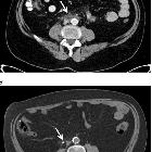
Retroperitoneal fibrosis is visible as a soft tissue density mass located around the aorta and iliac arteries. Classically, it develops around the aortic bifurcation and spreads upwards where it can envelop the renal hila. It encases but does not invade or stenose the ureters or vessels. However, ureteric obstruction and venous thromboses can occur.
In early or active stages, variable enhancement can be seen with intravenous contrast while no enhancement may be seen in the quiescent disease.
MRI
MRI has been reported to be as sensitive as CT in its assessment of retroperitoneal fibrosis with the added advantage of high contrast resolution between closely apposed retroperitoneal structures. It can evaluate the urinary tracts using fast T2 weighted spin-echo sequences without requiring intravenous contrast in patients with impaired renal function.
The soft tissue mass is usually dark on T1W and T2W unless there is an active inflammation whereby the T2W images can be hyperintense.
FDG-PET
Uptake will be seen in active inflammation and absent in metabolically inactive disease .
Treatment and prognosis
Retroperitoneal fibrosis may lead to ureteric obstruction and consequent renal failure. In some cases, a blow-out of the ureter is described as a result of the obstruction.
Differential diagnosis
Imaging differential considerations include:
- retroperitoneal lymphoma: non-lymphomatous RPF is often seen centered at L4/5 level; if the disease is centered cranially to L4/5, consider lymphoma
- retroperitoneal Erdheim-Chester disease
- retroperitoneal extramedullary hematopoiesis (rare)
Siehe auch:
- Morbus Ormond
- retroperitoneales Hämatom
- retroperitoneale Tumoren
- inflammatory abdominal aortic aneurysm
- Retroperitonealfibrose nach EVAR
- primäre intestinale Lymphangiektasie
- chronische Periaortitis
und weiter:
- genitourinary curriculum
- inflammatorischer Pseudotumor der Orbita
- Lymphom der Niere
- Zöliakusblockade
- Schilddrüse
- urinoma
- fibrosierende Mediastinitis
- IgG4-assoziierte Erkrankung
- Riedel-Thyreoiditis
- penetrating aortic ulcer and retroperitoneal fibrosis
- Obstruktion Vena cava inferior
- MR Imaging of perirenal idiopathic retroperitoneal fibrosis
- IgG4 assozierte retroperitoneale Fibrose
- perirenale Fibromatose

 Assoziationen und Differentialdiagnosen zu retroperitoneale Fibrose allgemein:


