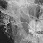
salt and pepper sign


The salt and pepper sign is used to refer to a speckled appearance of tissue. It is used in many instances, but most commonly on MRI. NB: pathologists also use the term.
Differential diagnosis
Vascular tumors
Used to describe some highly vascular tumors which contain foci of hemorrhage, typically a paraganglioma. The appearance is on T1 weighted sequences, and is made up of:
- punctate regions of hyperintensity = salt
- small flow voids = pepper
Typical tumors with this appearance include:
- paragangliomas
Similar appearance may be seen on T2 sequences or post contrast T1 sequences in patients with juvenile nasopharyngeal angiofibromas .
This appearance can also be seen in other hypervascular tumors such as metastatic hypernephroma or metastatic thyroid carcinoma .
Sjögren syndrome
The parotid gland in Sjögren syndrome has also been described as having a salt and pepper appearance, due to a combination of punctate regions of calcification (pepper) and fatty replacement (salt)
Vertebral hemangioma
A less common usage for the term is for vertebral hemangiomas which have a coarser black and white dotted appearance especially on axial T2 and T1 images (salt = fat, pepper = coarsened trabeculae).
Autosomal recessive polycystic kidney disease
The heterogeneous echotexture of the enlarged kidneys in autosomal recessive polycystic kidney disease has also been described as salt and pepper pattern .
MRI noise artefact
A related use of the term is to describe the noise sometimes seen in MRI images.
When you think about it, you can probably find folk who have used the term for all sorts of lesions; any lesion that has a fine granular imaging texture will do the trick.
See Also
- salt and pepper sign in the skull (pepper pot skull)
- raindrop skull
Siehe auch:

 Assoziationen und Differentialdiagnosen zu Pfeffer und Salz Erscheinungsbild:


