Carotid body tumor


Carotid body tumor, also known as a chemodectoma or carotid body paraganglioma, is a highly vascular glomus tumor that arises from the paraganglion cells of the carotid body. It is located at the carotid bifurcation with characteristic splaying of the ICA and ECA.
Epidemiology
Typically, carotid body tumors are diagnosed in the 4 to 5 decades, and have a female predilection like the other paragangliomas of the head and neck . They are the most common type of paraganglioma of the head and neck (account for 60-70%). In approximately 10% of cases, they are bilateral .
A small number are familial (7-10%), and in such cases, they are frequently multicentric (35-50%) . When familial, they are usually autosomal dominant in inheritance, and associated with :
Clinical presentation
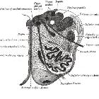
Clinical presentation is usually with a slow growing rounded neck mass. It is usually located anterior to the sternocleidomastoid near the angle of the mandible at the level of the hyoid bone. Characteristically, the tumor can be moved side to side but not up or down, due to its location within the carotid sheath .
Cranial nerves that travel in the carotid sheath (glossopharyngeal, vagus, accessory and hypoglossal nerves) may be involved. Associated symptoms relate to their dysfunction .
These tumor may synthesize and secrete catecholamines, although this is less common than with adrenal paragangliomas (pheochromocytomas) .
Pathology
The paraganglioma article includes a general discussion of the pathology of these tumors.
Radiographic features
Carotid body tumors are located at the carotid bifurcation with characteristic splaying of the ICA and ECA, described as the lyre sign. In all modalities, the dense vascularity of these tumors is manifested as prominent contrast enhancement.
CT
Contrast-enhanced CT is excellent at depicting these lesions. Typical appearances are:
- soft tissue density on non-contrast CT (similar to muscle)
- bright and rapid (faster than schwannoma) enhancement
- splaying of the ICA and ECA
- circumferential angle of contact of tumor with ICA could also be categorized under the Shamblin group system as
- group I: <180 degrees of encasement
- group II: 180-270 degrees of encasement
- group III: >270 degrees of encasement
- helps in deciding the risk of ICA adventitial involvement and possible need of ICA resection followed by grafting required in group III cases
MRI
- T1
- iso to hypointense compared to muscle
- salt and pepper appearance when larger, representing a combination of punctate regions of hemorrhage or slow flow (salt) and flow voids (pepper)
- intense enhancement following gadolinium
- T2
- hyperintense compared to muscle
- salt and pepper appearance also seen on T2
DSA/angiography
The splaying of the carotid vessels (lyre sign) is again identified with an intense blush in tumor with and 'early vein' seen due to arteriovenous shunting .
The ascending pharyngeal artery is the main contributing supply.
Scintigraphy
Although not specific, shows uptake with metaiodobenzylguanidine (MIBG) and octreoscan scintigraphy and can be useful for assessing multiple lesions.
Treatment and prognosis
Surgical excision is the treatment of choice. The larger the tumor the higher the risk of operative complications . In patients for whom the risk of complications precludes surgery, radiotherapy may be considered .
Malignant transformation is encountered in 2-36% of cases with metastases most commonly to bone, lung and liver and regional lymph nodes .
Differential diagnosis
General imaging differential considerations include:
- vagal schwannoma: spreads the ICA and internal jugular vein; displaces the ICA anteromedially
- vagal neurofibroma
- lymph node mass: may look similar if hypervascular
- glomus vagale tumor: same pathology but located more rostrally
- carotid bulb ectasia
Siehe auch:
- Arteria carotis externa
- Arteria carotis interna
- Arteria carotis communis
- Medulla oblongata
- carotid bifurcation
- Glomus caroticum
- Paragangliom des Nervus vagus
- Paragangliome von Kopf und Hals
- Klassifikation Glomus caroticum Tumor
- Klassifikation nach Shamblin Glomus caroticum Tumor
- Klassifikation nach Linder Glomus caroticum Tumor
und weiter:


 Assoziationen und Differentialdiagnosen zu Glomus caroticum Tumor: