Sichelzellenanämie (abdominelle Manifestationen)

Computed
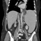
tomography of the spleen: how to interpret the hypodense lesion. Transverse contrast-enhanced CT images acquired during the portal venous phase. a A 43-year-old man with advanced sickle-cell disease. Note the irregular shape of the spleen, as well as the increased density of the splenic parenchyma, together with extensive calcifications as a consequence of constantly occurring micro-infarctions. b A 38-year-old man with sickle-cell disease exhibiting end-stage splenic involvement. Note the increased density, calcifications of the shrunken spleen

Imaging
findings of splenic emergencies: a pictorial review. Splenic sequestration. (a) CT features of ASSC. Contrast-enhanced axial CT image of a 26-year-old male with known sickle cell-thalassemia reveals lack of enhancement in spleen parenchyma (arrow). Retrograde filling of splenic vein (arrowhead) from the portal vein can be visualized. (b) Axial T2-weighted MRI demonstrates hypointense spleen (arrows) secondary to iron deposition. Splenic vein manifests with hyperintense appearance (arrowheads) due to slow flow resulting from retrograde venous filling

Sickle cell
disease • Sickle cell disease - Ganzer Fall bei Radiopaedia

Sickle cell
disease (abdominal manifestations) • Sickle cell disease (abdominal manifestations) - Ganzer Fall bei Radiopaedia

Sickle cell
disease • Sickle cell disease - Ganzer Fall bei Radiopaedia

Gallstones
• Cholelithiasis in sickle cell anemia - Ganzer Fall bei Radiopaedia
Abdominal manifestations of sickle cell disease (SCD) are wide and can involve many organs.
For a general discussion, please refer to sickle cell disease.
Splenic
- splenomegaly
- may occur transiently with the sequestration syndrome, where rapid pooling of blood occurs in the spleen, resulting in intravascular volume depletion, with potential for cardiovascular collapse
- autosplenectomy
- the slow, tortuous micro-circulation of the spleen renders it susceptible to infarction and subsequent functional asplenia
- 94% are asplenic by age 5
- radiological finding is of a small, calcified spleen
- splenic abscesses
Hepatobiliary
- hepatic iron deposition secondary to multiple transfusions
- hepatomegaly +/- coarsened echotexture with portal hypertension
- cholelithiasis +/- choledocholithiasis
- multiple liver abscesses
Renal
- kidneys are often large early in the disease, with variable echogenicity on ultrasound, but shrink with development of renal failure. Bilateral echogenic pyramids are frequently seen in sickle cell disease
- renal papillary necrosis
- renal vein thrombosis
Gastrointestinal tract
- approximately 40% patient may develop peptic ulcers due to reduced mucosal resistance and bowel ischemia
See also
Siehe auch:
- Splenomegalie
- Sichelzellenanämie
- portale Hypertension
- Autosplenektomie
- Papillennekrose der Niere
- muskuloskelettale Manifestationen bei Sichelzellanämie
- Acute chest syndrome bei Sichelzellanämie
- Gallensteine bei Kindern
- Sichelzellenanämie (zerebrale Manifestationen)
- Cholelithiasis bei Sichelzellenanämie
- Sichelzellenanämie (chronische Lungenerkrankung)
- coarsened echotexture


 Assoziationen und Differentialdiagnosen zu Sichelzellenanämie (abdominelle Manifestationen):
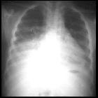


muskuloskelettale
Manifestationen bei Sichelzellanämie
