sickle cell spine

Young adult
with back pain. Lateral radiograph of the thoracic spine shows multiple H-shaped vertebral bodies.The diagnosis was sickle cell disease.

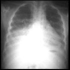
Teenager with
fever and sickle cell disease. CXR AP (left) shows a dense opacity in the left lower lobe which on the lateral (right) is seen to be located posteriorly, causing a spine sign. The lateral radiograph also shows the T8 and T10 vertebral bodes to be H-shaped.The diagnosis was acute chest syndrome and musculoskeletal manifestations of sickle cell disease.

Sickle cell
disease • Sickle cell disease with osseous changes and splenomegaly - Ganzer Fall bei Radiopaedia

Sickle cell
disease • H-shaped vertebrae - Ganzer Fall bei Radiopaedia

Sickle cell
disease • Sickle cell disease - Ganzer Fall bei Radiopaedia

Sickle cell
disease • H-shaped vertebrae due to sickle cell disease - Ganzer Fall bei Radiopaedia

Sickle cell
disease • Sickle cell disease - skeletal - Ganzer Fall bei Radiopaedia

Sickle cell
disease • Sickle cell disease - Ganzer Fall bei Radiopaedia

Teenager with
sickle cell disease and respiratory distress. CXR AP (left) shows opacity in the left lower lobe that on the lateral (middle) is located posteriorly (spine sign). The vertebral bodies (right) have an H-shaped appearance throughout the thoracic and lumbar spine.The diagnosis was acute chest syndrome and musculoskeletal manifestations of sickle cell disease.
Sichelzellenanämie Wirbelsäule
sickle cell spine
muskuloskelettale Manifestationen bei Sichelzellanämie Radiopaedia • CC-by-nc-sa 3.0 • de
Skeletal manifestations of sickle cell disease result from three interconnected sequelae of sickle cell disease :
These, in turn, can predispose individuals to other complications, such as growth disturbance and pathological fractures.
For a general discussion of sickle cell disease, please refer to sickle cell disease.
Clinical presentation
Vaso-occlusive crises are common and usually result in skeletal pain. Osteomyelitis will present with localized pain and systemic features of infection.
Radiographic features
The radiographic features will depend on the specific complication and are therefore discussed separately were appropriate.
- vaso-occlusive crises
- osteonecrosis
- bone infarcts typically involve medullary cavities and epiphyses
- the proximal humeri, proximal femora, and vertebral bodies are often affected
- in the humeri, serpiginous sclerosis is characteristic of infarction
- vertebral infarcts may result in
- H-shaped vertebrae
- tower vertebra
- vanishing vertebra
- anterior vertebral vascular notches
- subperiosteal and epidural hemorrhage
- hand-foot syndrome (dactylitis)
- imaging findings include patchy areas of lucency with periostitis and soft tissue swelling of metacarpals or metatarsals which are difficult to distinguish from osteomyelitis
- growth disturbance
- osteonecrosis
- chronic anemia
- marrow hyperplasia
- long bones: widening of medullary spaces and thinning of cortical bone
- skull: widening of diploic space, thinning of the outer table, hair-on-end appearance
- osteopenia and pathological fractures
- extramedullary hematopoiesis
- red marrow reconversion
- marrow hyperplasia
- infection
See also
Siehe auch:

 Assoziationen und Differentialdiagnosen zu Sichelzellenanämie Wirbelsäule:
muskuloskelettale
Manifestationen bei Sichelzellanämie


