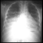
Sickle cell disease (acute chest syndrome)


Acute chest syndrome (ACS) in sickle cell disease is a leading thoracic complication - as well as leading cause of mortality - in those affected by sickle cell disease. The diagnosis is made on the combination of new pulmonary opacity on chest x-ray with at least one new clinical symptom or sign.
For a general discussion of sickle cell disease, please refer to sickle cell disease.
Clinical presentation
Patients may present with acute fever, cough, wheezing, tachypnea and/or chest pain on a background of established sickle cell disease.
Pathology
There is no single underlying etiology to acute chest syndrome but rather a variety of infectious and noninfectious causes, including :
- pneumonia
- pulmonary infarction
- fat embolism
- rib or sternal infarct causing atelectasis (from splinting)
Radiographic features
Plain radiograph
Typically seen as segmental or subsegmental atelectasis/consolidation with a lower lobe predilection, and/or pleural effusion.
A chest radiograph may also show other sequelae from sickle cell disease such as bone infarcts, rib enlargement and cardiomegaly (from anemia).
Ultrasound
Point-of-care lung ultrasonography in the acute chest syndrome may reveal one of the following patterns;
- alveolar consolidation
- the most common abnormality found, with a posterobasal regional predilection
- air bronchograms may be visualized
- anterior subpleural consolidations
- presence of small (≤1 cm) crescentic hypoechoic collections disrupting the pleural line (C-lines thought to represent lung infarction )
- often associated with adjacent pleural thickening and loss of lung sliding
- lung rockets
- three or more B-lines per sonographic field, typically 3 cm apart (B3 lines) - defines the sonographic interstitial syndrome
- bilateral diffuse anterolateral interstitial syndrome may be observed
- pleural effusions
CT
May show a mosaic perfusion pattern that could be associated with a pleural effusion. The radiographic signs above may also be seen on CT.
Differential diagnosis
General imaging differential considerations include:
See also
Siehe auch:

 Assoziationen und Differentialdiagnosen zu Acute chest syndrome bei Sichelzellanämie:


