spinal arteriovenous malformation


Spinal arteriovenous malformations (AVMs) are characterized by arteriovenous shunting with a true nidus. They represent ~25% of spinal vascular malformations.
Epidemiology
Different types of spinal AVM (see below) have differing age of presentation, but overall 80% present between the age 20 and 60 years .
Clinical presentation
It is variable, ranging from the progressive myelopathy (Foix-Alajouanine syndrome), often with delayed diagnosis, to the catastrophic spinal subarachnoid hemorrhage (see: coup de poignard of Michon ).
Pathology
Classification
Spinal AVMs may be classified as intramedullary and extramedullary (80%) and further divided into four angiographic types, with additional subtypes (see: spinal AVM classification).
Complications
- myelopathy from venous congestion/hypertension
- hemorrhage: within the cord parenchyma or subarachnoid space
- high-flow AVMs may cause arterial steal from adjacent spinal cord segments
- myelopathy from large AVMs (rare)
Radiographic features
Digital subtraction angiography (DSA)
Angiography remains the investigation of choice but requires meticulous technique. It is essential to remember that the site of arterial supply can be anywhere from the upper thoracic to sacral areas with little relationship to the clinical level, or visible nidus (if present).
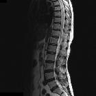
MRI
Signal characteristics
- T1
- signal voids from high-velocity flow
- dilated perimedullary vessels indent/scallop the cord
- T2
- signal voids from high-velocity flow
- increased cord signal due to cytotoxic edema or myelomalacia
Treatment and prognosis
Both surgery and angioembolisation have a role in the treatment of spinal AVMs .
Siehe auch:
- Kaudaredundanz
- Arteriovenöse Malformation
- Subarachnoidalblutung
- spinale durale arteriovenöse Fistel
- spinal AVM classification
- coup de poignard of Michon
- Myelopathia necrotisans
und weiter:


 Assoziationen und Differentialdiagnosen zu spinale arteriovenöse Malformationen: