tricuspid atresia


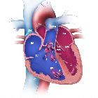
Tricuspid atresia is a cyanotic congenital cardiac anomaly which is characterized by agenesis of the tricuspid valve and right ventricular inlet. There is almost always an obligatory intra-atrial connection through either an ASD or patent foramen ovale (PFO) in order for circulation to be complete . A small VSD is often also present. In a proportion of cases, tricuspid atresia may also be associated with transposition of great arteries (TGA).
Clinical presentation
ECG
- giant (>5 mm) P waves
- with peaked morphology in lead II
- exaggeration of right atrial enlargement morphology
- referred to as "Himalayan P waves"
- extreme axis deviation
- high left ventricular voltage (HLVV)
- left ventricular hypertrophy voltage criteria
Pathology
It results from an unequal atrioventricular canal division and the right ventricle is typically very hypoplastic.
Associations
Recognized extra-cardiac associations include:
- right-sided aortic arch
- absent spleen: asplenia
Radiographic features
Plain radiograph
Chest radiographic features may vary depending on the presence and extent of a VSD or TGA. May demonstrate decreased pulmonary vascularity (i.e. oligaemic appearance). Cardiac size may be normal or enlarged.
Echocardiography/ultrasound
Usually the 1st line imaging modality in utero. It allows direct visualization of the anomaly.
CT and MRI
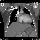
Allows direct visualization of the anomaly and may typically show a fatty and/or muscular separation of the right atrium from the right ventricle. Cine MRI can offer functional information in addition to anatomy.
Siehe auch:
- Atriumseptumdefekt
- rechts descendierende Aorta
- Transposition der großen Arterien
- Ventrikelseptumdefekt
- Single Ventricle
- Trikuspidalklappenstenose
- cyanotic congenital cardiac anomaly
und weiter:

 Assoziationen und Differentialdiagnosen zu tricuspid atresia:


