Trikuspidalklappenstenose
Tricuspid valve stenosis is a valvulopathy that describes narrowing of the opening of the tricuspid valve between the right ventricle and the right atrium.
Epidemiology
MS is seen more commonly in women and in countries, generally developing nations, where rheumatic fever is common .
Clinical presentation
Patients with tricuspid stenosis characteristically present with right-predominant clinical features of heart failure such as hepatomegaly, ascites, and anasarca . Clinical examination classically reveals an elevated jugular venous pressure with a dominant a-wave and slow y-descent, and a mid-diastolic murmur that is heard on praecordial auscultation .
Tricuspid stenosis rarely occurs in isolation, and usually presents when there is already significant mitral stenosis in the setting of rheumatic heart disease . However, patients with these dual valvulopathies rarely present with the left-predominent clinical features of heart failure because the tricuspid stenosis prevents blood from entering the pulmonary circulation . Thus, somewhat paradoxically, the degree of dyspnea may be minimal compared to the degree to tricuspid stenosis .
Pathology
Tricuspid stenosis is usually acquired via rheumatic heart disease, where there is chronic inflammation of the tricuspid valve leaflets (tricuspid valvulitis) leading to eventual thickening, calcification, immobilization, and narrowing of the tricuspid valve . This occurs in a similar manner to rheumatic mitral stenosis (see individual article for detailed discussion of this pathophysiology).
The characteristic hemodynamic feature of tricuspid stenosis is an increased diastolic pressure gradient between the right atrium and right ventricle . A pressure ≥5 mmHg is significant enough to result in venous congestion, explaining many of the clinical features of tricuspid stenosis . Eventually, right atrial enlargement manifests, cardiac output decreases, and often tricuspid regurgitation also occurs .
It is important to differentiate tricuspid stenosis from tricuspid hypoplasia, where in the latter, the right ventricle also often tends to also be hypoplastic.
Etiology
In addition to being a sequela of rheumatic fever, which is by far the most common cause world-wide, there are other rarer causes :
- congenital tricuspid stenosis
- carcinoid heart disease (always combined with tricuspid regurgitation)
- nonbacterial thrombotic endocarditis
- infective endocarditis
- fibrosis/adhesions associated with endocardial pacemaker leads
- Fabry disease
- cardiac amyloidosis
- Whipple disease
- right atrial myxoma (generally not considered 'true' tricuspid stenosis)
In these non-rheumatic causes, tricuspid stenosis can occur in isolation without any mitral valve disease .
Radiographic features
Plain radiograph
Signs of tricuspid stenosis on chest radiograph are often subtle, especially due to the presence of signs of concurrent mitral stenosis, but include :
- right atrial enlargement
- superior vena caval enlargement
- features of mitral stenosis
- rarely, calcifications of the tricuspid valve may be seen
- features of congestive heart failure may also be present
Ultrasound: echocardiography
Echocardiography is useful for assessing the tricuspid valve area, jet velocity, pressure gradients, and the right atrium. Two-dimensional echocardiographic features which may suggest the diagnosis include:
- thickened, echogenic valve leaflets
- the perivalvular apparatus should be carefully assessed for an atrial mass, or the presence of vegetations
- impaired leaflet excursion with diastolic doming
- marked restriction of leaflet separation common in carcinoid syndrome
- right atrial enlargement
- inferior vena cava plethora
Hemodynamic parameters, calculated by spectral Doppler interrogation of transvalvular flow, used to determine what constitutes significant tricuspid stenosis include :
- mean pressure gradient ≥5 mmHg
- trans-tricuspid velocity time integral >60 cm
- pressure half time ≥190 ms
- derived valve area ≤1 cm
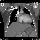
CT/MRI
Cross-sectional imaging demonstrate the same radiographic features appreciated on plain film and echocardiography, but in greater detail . In particular, cardiac MRI may be particularly useful for accurate measurements to assess the severity of the valvulopathy .
Treatment and prognosis
The decision to treat tricuspid stenosis is based on its severity. Management involves pharmacotherapy measures (especially diuretics) and consideration of surgery . Surgery is generally recommended at the same time as mitral valve surgery for mitral stenosis . Details of this management is beyond the scope of this article.
Complications
See also
- valvular heart disease
- general tricuspid valve pathologies:
- tricuspid valve stenosis
- tricuspid valve regurgitation
- specific tricuspid valve pathologies:
- congenital tricuspid valve stenosis
- Ebstein anomaly
- tricuspid valve atresia
Siehe auch:
und weiter:


 Assoziationen und Differentialdiagnosen zu Trikuspidalklappenstenose: