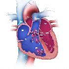
Transposition der großen Arterien


 nicht verwechseln mit: TGA transiente globale Amnesie
Transposition of the great arteries (TGA) (also known as transposition of the great vessels (TGV)) is the most common cyanotic congenital cardiac anomaly presenting during the newborn period, with cyanosis in the first 24 hours of life. It accounts for up to 7% of all congenital cardiac anomalies and can be assessed with echocardiography, gated cardiac CT, or cardiac MRI.
Epidemiology
The estimated incidence is at ~1 in 5000 births. Transposition of the great arteries is an isolated abnormality in 90% of those affected and rarely is associated with a syndrome or an extra-cardiac malformation. It is most common in infants of diabetic mothers .
Pathology
Occurs as a result of ventriculoarterial discordance, with the aorta arising from the right ventricle and the pulmonary trunk from the left ventricle. It can be subdivided into two main types depending on the positional relationship of the aortic valve with the pulmonary valve:
- L-loop transposition of the great arteries: congenitally corrected TGA
- D-loop transposition of the great arteries
This article mainly focuses on the D-loop transposition.
An isolated TGA is incompatible with life at birth without one of the following additional anomalies (which are a common occurrence ):
- atrial septal defect (ASD): uncommon
- ventricular septal defect (VSD): ~35%
- patent ductus arteriosus (PDA): unstable due closure following birth
- patent foramen ovale (PFO): unstable
Unstable associations account for 60-65% of occurrences.
Associations
Approximately 90% of TGAs occur as an isolated finding, and extracardiac syndromic associations are rare. Associations have been described with:
- maternal diabetes
- congenital coronary arterial anomalies
Radiographic features
Plain radiograph
A frontal chest radiograph classically shows cardiomegaly with cardiac contours classically described as appearing like an egg on string . There is often an apparent narrowing of the superior mediastinum as the result of the aortic and pulmonary arterial configuration, i.e. parallel in D-loop transposition, with the main pulmonary artery posterior to the aorta.
Echocardiography
Allows direct visualization of abnormal anatomy with the aorta and pulmonary trunk lying in parallel with an absence of crossing (best seen in the base view of the fetal heart) .
CT/CTA
Allows direct visualization of abnormal great vessel anatomy. Cardiac-gated cine CT can additionally assess function.
Cardiac MRI
Allows direct visualization of abnormal anatomy. SSFP cine sequences can additionally determine flow dynamics.
Treatment and prognosis
Initial Rashkind septoplasty is usually done as a palliative procedure in neonates.
Definitive surgical correction
Previously TGAs were treated with atrial switch operations, such as a Mustard repair or Senning repair, which have been superseded by arterial switch procedures .
Siehe auch:
- Persistierender Ductus arteriosus
- Atriumseptumdefekt
- Herzfehler
- rechts descendierende Aorta
- transiente globale Amnesie
- Ventrikelseptumdefekt
- Mustard repair
- SSFP - steady state free precession
- TGA
- cyanotic congenital cardiac anomalies
und weiter:
- Situs inversus
- Varianten der Herzanatomie
- fetal conditions associated with maternal diabetes
- tricuspid atresia
- CXR approach to congenital heart disease
- chest x-ray appeoach to congenital heart disease
- great vessels
- congenital heart disease - chest x-ray approach
- Zyanotischer Herzfehler
- isolated unilateral absence pulmonary artery (IUAPA)
- egg on a string
- magnetic resonance imaging of congenitally corrected transposition of the great arteries

 Assoziationen und Differentialdiagnosen zu Transposition der großen Arterien:


