ventricular septal defect (VSD)

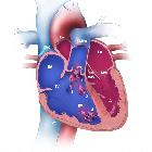


Ventricular septal defects (VSD) represent defects in the interventricular septum that allow a hemodynamic communication between the right and left ventricles. It typically results in a left-to-right shunt.
Epidemiology
They represent one of the most common congenital cardiac anomalies and may be associated with up to 40% of such anomalies . They are considered the most common congenital cardiac abnormality diagnosed in children and the second most common diagnosis in adults. The estimated incidence is at ~1 in 400 births .
Clinical presentation
Clinical presentation varies depending on the size and resultant severity of the VSD . Small lesions with minimal shunting may be asymptomatic, however may have a loud harsh pansystolic murmur heard on precordial auscultation over the left sternal border . Larger lesions, in comparison, may cause signs of heart failure such as exertional dyspnea, raised jugular venous pressure, hepatomegaly, peripheral edema, or failure to thrive in pediatric patients, but may have a very soft murmur .
In addition to a pansystolic murmur, there may also be a mid/end-diastolic murmur mimicking mitral stenosis as well as an ejection systolic murmur mimicking aortic stenosis, each due to increased pulmonary circulation compared to systemic circulation resulting in more total blood passing through the left atrium and ventricle . Cyanosis is generally not seen in patients with VSDs (unless Eisenmenger syndrome develops).
ECG
While small VSDs will remain electrocardiographically occult, larger defects will classically demonstrate some of the following features :
- left atrial enlargement
- high left ventricular voltage (HLVV)
- voltage criteria for left ventricular hypertrophy
- prominent, biphasic RS complexes (V2-V4) with amplitudes > 50 mm
- referred to as the Katz-Wachtel phenomenon
Pathology
Classification according to location
- membranous/perimembranous (most common: 80-90%)
- including the Gerbode defect
- inlet/inflow
- outlet/subarterial
- muscular/trabecular
Associations
A VSD can occur on its own but frequently tends to occur with other cardiovascular associations:
- cardiovascular associations
- tetralogy of Fallot
- truncus arteriosus
- double outlet right ventricle: including Taussig-Bing malformation
- aortic coarctation
- tricuspid atresia
- aortic regurgitation
- pulmonary stenosis
- extra-cardiac associations
- aneuploidic/chromosomal anomalies
- other syndromic anomalies: there are several and include
Radiographic features
Plain radiograph
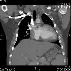
The chest radiograph can be normal with a small VSD. Larger VSDs may show cardiomegaly (particularly left atrial enlargement although the right and left ventricle can also be enlarged). A large VSD may also show features of pulmonary arterial hypertension, pulmonary edema, pleural effusion, and increased pulmonary vascular markings.
Ultrasound: echocardiography
Allows direct visualization of the septal defect; a transthoracic parasternal short axis at the level of the aortic valve is typically the view of choice for differentiation between supracristal and perimembranous defects, whereas apical and subcostal windows are preferred for muscular defects .
Specific features sought after include:
- defect localization
- a perimembranous VSD can be seen as a septal dropout in the area adjacent to the tricuspid septal leaflet and below the right border of the aortic annulus
- muscular VSDs are variably located along the septum
- may be numerous
- inlet VSDs involve the septal component of the right ventricular inflow tract
- supracristal VSDs are visualized as dropout above the crista supraventricularis in the right ventricular outflow tract
- often associated with aortic regurgitation
- direction of VSD jet
- a parasternal short axis at the aortic valve level will reveal aliased flow at the 10 o'clock position in a perimembranous defect
- a supracristal VSD will demonstrate flow at the 2 o'clock position
- hemodynamic consequences
- restrictive VSD's have a more benign prognosis
- tend to be smaller defects with a large attendant pressure gradient
- rarely progress to cause structural or hemodynamic derangement
- non-restrictive septal defects are larger and tend to transmit significant volume to the right-sided circulation
- demonstrate low-velocity jets with a much lower pressure gradient
- associated with dilation of the left ventricle and pulmonary hypertension
- the pulmonary artery systolic pressure (sPAP) may be calculated using the doppler acquired velocity across the defect and the systolic blood pressure
- sPAP = SBP - 4(jet velocity)^2
- the pulmonary artery systolic pressure (sPAP) may be calculated using the doppler acquired velocity across the defect and the systolic blood pressure
- restrictive VSD's have a more benign prognosis
CT
CTA with ECG-gating allows direct visualization of the defect. Large VSD's may be seen on non-gated studies.
MRI
May also show added functional information (e.g. quantification/shunt severity) in addition to anatomy. Some muscular defects can give a "Swiss cheese" appearance owing to their complexity.
Treatment and prognosis
The prognosis is good for small VSDs which show a high spontaneous intrauterine or postnatal closure rate. VSDs usually do not cause any hemodynamic compromise in utero due to the right and left ventricular pressures being very similar during that period.
Complications
- Eisenmenger phenomenon with shunt reversal (i.e. left-to-right becomes right-to-left)
- cardiac failure
Siehe auch:
- Aortenisthmusstenose
- Atriumseptumdefekt
- Pätau-Syndrom
- Down-Syndrom
- Fallot'sche Tetralogie
- Trisomie 18
- tricuspid atresia
- double outlet right ventricle (DORV)
- chromosomale Anomalien
- Holt-Oram-Syndrom
- Eisenmenger-Reaktion
- Pulmonalstenose
- Truncus arteriosus communis
- aneuploidic
und weiter:
- Ektasie Aorta ascendens
- Herzfehler
- Meckel-Syndrom
- Kardiomegalie
- acyanotic congenital heart disease
- Varianten der Herzanatomie
- Transposition der großen Arterien
- Trisomie 22
- fetal conditions associated with maternal diabetes
- vergößerter linker Vorhof
- Bland-White-Garland-Syndrom
- causes of pulmonary arterial hypertension
- antenatal features of Down syndrome
- CXR approach to congenital heart disease
- Embryopathia rubeolosa
- quadricuspid aortic valve
- Thalidomid-Embryopathie
- Rubinstein-Taybi-Syndrom
- tetralogy of fallot (mnemonic)
- fetal right ventricular enlargement
- Fetales Alkoholsyndrom
- chest x-ray appeoach to congenital heart disease
- dysencephalia spanchnocystica
- congenital heart disease - chest x-ray approach
- Cantrell’sche Pentalogie
- pentalogy of Fallot
- anomalous left coronary artery off the pulmonary artery
- four chamber cardiac view
- isolated unilateral absence pulmonary artery (IUAPA)
- differential of an enlarged pulmonary trunk on chest radiography
- erweiterte Pulmonalarterie
- Herzektopie
- Nicht-zyanotische Herzfehler
- pulmonary atresia with ventricular septal defect and situs inversus totalis
- apical ventricular septal defect
- post-infarction ventricular septal defect


 Assoziationen und Differentialdiagnosen zu Ventrikelseptumdefekt: