tricuspid insufficiency


Tricuspid valve regurgitation (TR), also known as tricuspid valve insufficiency or tricuspid valve incompetence (TI), is a valvulopathy that describes leaking of the tricuspid valve (TV) during systole that causes blood to flow in the reverse direction from the right ventricle (RV) into the right atrium (RA).
Epidemiology
The prevalence of moderate or severe tricuspid regurgitation is 0.8%, with this prevalence increasing with aging . Women are 4.3 times more likely to be affected .
Clinical presentation
Clinical features can vary dependent on the severity of the tricuspid regurgitation, and often only manifest when tricuspid regurgitation is severe . When symptomatic, it eventually leads to right-predominant clinical features of heart failure and clinical features of pulmonary hypertension .
Clinical examination classically reveals an elevated jugular venous pressure with dominant c-v-waves and a sharp y-descent, pulsatile hepatomegaly, and a pansystolic (holosystolic) murmur that is heard on precordial auscultation that becomes louder on inspiration (Carvallo sign) .
Pathology
Hemodynamic consequences of tricuspid regurgitation only become apparent in severe TR . This is because the right atrium is very compliant and is able to accommodate for reasonably large regurgitant volumes . Eventually, as tricuspid regurgitation becomes more severe, the right atrial and venous pressures will eventually increase, leading to the classic signs and symptoms, and there is a decrease in forward cardiac output .
Etiology
In a majority of patients (70-85%), the tricuspid regurgitation is considered 'functional' (or 'secondary'), where it is caused by dilatation of the annulus as a result of increased pulmonary and right ventricular pressures . In the remaining 15–30% of patients, it may be organic (or 'primary') and related to direct involvement of the tricuspid valve . Furthermore, tricuspid regurgitation in isolation is very rare; it is more often found in association with other valvular disease, especially mitral valve disease .
Causes of organic (primary) tricuspid regurgitation include :
- congenital abnormalities (e.g. Ebstein anomaly)
- direct factors affecting the valve
- myxomatous degeneration (associated with mitral valve prolapse)
- infective endocarditis
- rheumatic heart disease
- valve dysfunction due to myocardial infarction
- valve disruption from trauma
- carcinoid heart disease
- connective tissue disorders (e.g. Marfan syndrome)
- drug-induced disease (e.g. exposure to phentermine or fenfluramine)
Disorders leading to functional (secondary) tricuspid regurgitation include :
- causes of left-sided heart failure (especially from mitral stenosis or mitral regurgitation)
- pulmonary hypertension
- right ventricular myocardial infarction
- left-to-right shunts
- Eisenmenger syndrome
- pulmonary stenosis
- pulmonary regurgitation
- hyperthyroidism
Radiographic features
Plain radiograph
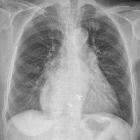
Signs of tricuspid regurgitation on chest radiograph are often subtle, but include :
- right atrial enlargement
- right ventricular enlargement
- reduced prominence of pulmonary vascularity
- superior vena caval enlargement
- inferior vena caval enlargement
- distension of azygos vein
- features of congestive heart failure may also be present
Ultrasound: echocardiography
Echocardiography is useful for evaluating the cause of tricuspid regurgitation, for assessing the regurgitant volume, and for assessing the right-sided cardiac chambers. It may be best evaluated from the apical window, however, left parasternal, right ventricular inlet view and short axis at the aortic valve level may be other useful positions. Hepatic vein Doppler is also often performed in the work-up for tricuspid regurgitation.
The tricuspid valve may be consistently visualized in the right ventricle modified parasternal long axis, apical 4 chamber, and right ventricular inflow-outflow views; only the modified parasternal long axis allows visualization of the posterior valve leaflet. Apical views
The echocardiographic detection of tricuspid regurgitation is common and present in approximately 70% of people. For pathological tricuspid regurgitation, various parameters are used in order to determine severity, such as :
- mild
- tricuspid valve normal
- right ventricular or right atrial or inferior vena caval size normal
- central jet <5 cm
- regurgitant volume not defined
- vena contracta not defined
- proximal isovelocity surface area radius ≤0.5 cm
- jet density and contour is soft and parabolic
- hepatic vein flow has systolic dominance
- moderate
- tricuspid valve may be normal or abnormal
- right ventricular or right atrial or inferior vena caval size may be normal or abnormal
- central jet 5-10 cm
- regurgitant volume not defined
- vena contracta not defined but <0.7 cm
- proximal isovelocity surface area radius 0.6-0.9 cm
- jet density and contour is dense with a variable contour
- hepatic vein flow has systolic blunting
- a pattern consisting of a pulsatile waveform with prominent v waves, decreased S wave amplitude, and reversal of the S/D ratio is specific for tricuspid regurgitation
- severe
- tricuspid valve is abnormal with a flail leaflet
- right ventricular or right atrial or inferior vena caval size usually dilated
- central jet >10 cm
- regurgitant volume ≥45 mL
- vena contracta >0.7 cm
- proximal isovelocity surface area radius >0.9 cm
- jet density and contour is dense and triangular with early peaking
- E-wave velocity >1 m/s
- hepatic vein flow has systolic reversal
- characteristic inversion of the S wave and fusion with the a and v waves will form a characteristic a-S-v complex
Additionally, the shape of the tricuspid annulus can alter with regurgitation . A normal annulus has a bimodal shape with distinct high points located anteroposteriorly and low points located mediolaterally . With functional tricuspid regurgitation, the annulus can become larger, more planar, and circular .
CT
CT can demonstrate the same radiographic features appreciated on plain film and echocardiography, but in greater detail .
Ancillary features
Dynamic contrast enhanced abdominal computed tomography (CT) may show intense opacification of the inferior vena cava and hepatic veins .
MRI
Cardiac MRI (CMR) is able to provide the most detailed assessment of the tricuspid valve and cardiac function . On spin-echo MR images, features that may be visible which could suggest towards a diagnosis of tricuspid regurgitation include :
- enlargement of the right ventricle
- enlargement of the right atrium
- distension of the venae cavae and hepatic veins
Cine gradient-echo imaging can be used to evaluate tricuspid regurgitation based on the area of the signal void corresponding to the regurgitant flow jet in systole . The signal void is best demonstrated on a four-chamber view and a coronal oblique view encompassing the right atrium and the right ventricle .
The degree of tricuspid regurgitation may be calculated in terms of regurgitant volume and fraction in similar ways to mitral regurgitation - i.e. subtraction of the forward stroke volume (as measured in the pulmonary artery with phase contrast) from the total right ventricular stroke volume (obtained from SSFP images) .
Treatment and prognosis
The decision to treat tricuspid regurgitation is based on the etiology and severity . Management involves pharmacotherapy measures (especially diuretics) and consideration of surgery . Surgery is generally recommended if there are signs of pulmonary hypertension, in which case tricuspid valve repair or tricuspid valve replacement may be necessary . Management of concurrent valvulopathies is also recommended . Details of this management are beyond the scope of this article.
Complications
See also
- valvular heart disease
- general tricuspid valve pathologies:
- tricuspid valve stenosis
- tricuspid valve regurgitation
- specific tricuspid valve pathologies:
- congenital tricuspid valve stenosis
- Ebstein anomaly
- tricuspid valve atresia
Siehe auch:
- TricValve® Transcatheter Bicaval Valves
- Trikuspidalklappenersatz
- Hedinger-Syndrom
- Karzinoidsyndrom
- Mitraclip an der Trikuspidalklappe
- Trikuspidalklappe
- Trikuspidalklappenanuloplastie
- Trikuspidalklappenstenose
- Mitralix Ltd. Mistral System device
- gastrointestinale neuroendokrine Tumoren
und weiter:

 Assoziationen und Differentialdiagnosen zu Trikuspidalklappeninsuffizienz:


