central nervous system vasculitides


Central nervous system (CNS) vasculitides represent a heterogeneous group of inflammatory diseases affecting the walls of blood vessels in the brain, spinal cord, and the meninges.
Please refer to the article on vasculitis for a general discussion of that entity.
The aim of this article will be to discuss the primary angiitis of the CNS (PACNS) since the other vasculitides are already discussed in specific articles.
Terminology
CNS vasculitides are classified as :
- primary: confined to the CNS with no involvement of other systems - referred to as primary angiitis of the CNS
- secondary: occurs in the context of a systemic inflammatory or infectious process
Please, note that this classification is different from that one used when discussing systemic vasculitides.
Epidemiology
Primary angiitis of the CNS remains a rare disorder: an estimated average annual incidence rate of 2.4 cases per million. It affects patients of all ages, but peaks at around 50 years of age, with males affected more commonly than females .
Secondary causes of CNS vasculitis far exceed the number of cases of primary angiitis of the CNS . Please refer to each specific vasculitis for further details.
Clinical presentation
Clinical features of primary angiitis of the CNS are non-specific. The diagnosis is made based on Calabrese’s criteria , including:
- the presence of an acquired otherwise unexplained neurological or psychiatric deficit
- the presence of either classic angiographic or histopathological features of angiitis within the CNS (biopsy remains the standard of reference for diagnosis )
- no evidence of systemic vasculitis or any disorder that could cause or mimic the angiographic or pathological features of the disease
When part of a systemic disorder, the diagnosis may be easier, unless the cerebral symptoms are the first to manifest. Please refer to a specific vasculitis for further details on clinical manifestation.
Pathology
For almost all forms of vasculitis, including primary angiitis of the CNS, the triggering factor is unknown .
CNS secondary vasculitides:
- affecting large blood vessels
- Takayasu arteritis: uncommon to have CNS involvement
- giant cell arteritis
- affecting medium blood vessels
- affecting small blood vessels
- IgA arteritis
- microscopic polyangiitis (microscopic polyarteritis)
- granulomatosis with polyangiitis
- eosinophilic granulomatosis with polyangiitis
- variable vessels sizes
- associated with systemic disease
- associated with a known etiology
- infection-induced vasculitis
- drug-induced
- malignant-induced
- radiation-induced
Radiographic features
Imaging findings for primary angiitis of the CNS are usually variable and non-specific, with ischemic infarctions the most common lesions, occurring in 53% of cases .
CT
May show areas of hypoattenuation.
Angiography
Digital subtraction angiography shows focal or multifocal segmental narrowing of both small and medium-sized blood vessels, occlusions may also be present. The same findings can be demonstrated in both CTA and MRA.
MRI
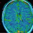
More specific to show multiple infarctions: usually bilateral, affecting different vascular territories of variable size, and in various stages of healing.
T2 and FLAIR high-intensity lesions in the white matter are also very common in primary angiitis of the CNS, but completely non-specific. Meningeal enhancement and intracranial hemorrhage can also be seen.
Vessel wall MRI (VW-MRI) may be a useful adjunct to conventional MRI, allowing differentiation between vasculitis, where there is contrast enhancement of the affected arterial wall, and other causes of vascular narrowing, such as intracranial atherosclerotic disease (ICAD) plaque, reversible cerebral vasoconstriction syndrome, dissection, and moyamoya disease .
Treatment and prognosis
Primary angiitis of the CNS is managed with high dose steroids and cytotoxic agents .
History and etymology
Primary angiitis of the CNS was initially reported in 1959 by Humberto Cravioto and Irwin Feigin .
Differential diagnosis
Practical points
Remember that despite being composed of non-specific findings, MRI is almost 100% sensitive for primary angiitis of the CNS and a normal MRI practically excludes this diagnosis .
Siehe auch:
- Churg-Strauss-Syndrom
- Vaskulitis
- zerebrale Mikroblutungen
- granulomatöse Angiitis des Zentralen Nervensystems
und weiter:


 Assoziationen und Differentialdiagnosen zu Vaskulitis des Zentralen Nervensystems:

