Coxarthrose


Osteoarthritis (OA) of the hip is the most common form of joint disorder of the hip, affecting primarily the articular cartilage of the hip joint and the surrounding tissues.
Epidemiology
The hip is the third most common joint affected by osteoarthritis after the knee and the hand . Women are more commonly affected than men. Reported prevalence varies in different studies and is also subject to geographic conditions. The lifetime risk of symptomatic hip osteoarthritis in people reaching the age of 85 years was estimated to be as high as 25% in certain regions .
Risk factors
Attributes, characteristics or exposures that increase the likelihood of developing osteoarthritis of the hip are :
- older age
- obesity
- genetics
- repetitive stress and mechanical overload
- farmers, construction workers
- high impact sports (football, handball, hockey, wrestling, weight-lifting, and long-distance running)
- acetabular dysplasia
- femoroacetabular impingement
- slipped capital femoral epiphysis
- Perthes disease
- trauma, e.g. hip dislocation or hip fracture
Clinical presentation
Patients usually experience slowly progressive hip pain, or hip-related groin pain radiating into the thigh, buttocks or knee. The pain can be worse at night, at rest or with strenuous activity, reducing the range of motion and limiting walking distance. It can be associated with stiffness particular in the morning or after rest. Other symptoms include locking, grinding and joint instability, fatigue and pain-related psychological stress .
Pathology
Osteoarthritis is characterised by an active progressive alteration of the whole synovial joint, due to a combination of mechanical, inflammatory and metabolic factors. This arises from an imbalance between destruction and repair of the affected tissues. The disease not only affects the hyaline cartilage, which loses its structural integrity due to composition changes but also involves the other tissues of the joint including the subchondral bone, the joint capsule and the synovium as well as the ligaments and the periarticular muscles .
Aetiology
- idiopathic/unknown
- previous trauma
- acetabular dysplasia
- femoroacetabular impingement
- inflammatory joint disease, e.g. septic arthritis
- haemochromatosis, haemophilia
- iatrogenic, e.g. multiple intra-articular steroid injections
Classification
Osteoarthritis of the hip can be classified into primary and secondary, depending on whether it is due to a known predisposing factor or not.
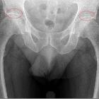
Radiographic features
General features are osteophyte formation, joint space narrowing and sclerosis of the subchondral bone plate. Subchondral cyst formation and remodelling of the articular surfaces or deformity are seen in more advanced stages.
Plain radiograph
Plain radiographs of the hip are a cheap, widely available and easily obtained modality and their interpretation in the evaluation of osteoarthritis is not as difficult as other imaging modalities .
For the indication of osteoarthritis of the hip, an anteroposterior radiograph of the hip and a cross-table lateral or frog-leg lateral view are obtained.
Hip joint space narrowing ≤2 mm or <2.5 mm or the combination of joint space narrowing with the presence of osteophytes, in particular, in the absence of any elevated inflammatory markers (e.g. ESR <20 mm/h) can be used as an indicator of osteoarthritis .
Radiological classification systems used for the assessment of osteoarthritis of the hip are the Kellgren and Lawrence score , the Croft score and the Tönnis classification, which are all susceptible to subjectivity, but the first being apparently the most reliable .
Another semi-quantitative method, which does not give a definition of osteoarthritis by grade, but grades different features of osteoarthritis as femoral and acetabular osteophyte formation as well as superior and medial joint space narrowing is the OARSI atlas .
Recommended scoring systems are the Kellgren and Lawrence score and the OARSI atlas .
Ultrasound
Ultrasound can depict joint effusions and synovitis by increased synovial vascularisation and detect osteophytes. In addition, it can be used for image-guided injections .
CT
Computed tomography provides information on the 3D assessment of acetabular and proximal femoral anatomy and can be used for surgical planning in cases of femoroacetabular impingement (FAI) or acetabular dysplasia or to assess the amount of bone stock .
MRI
In addition to the 3D visualisation of acetabular and femoral head-neck morphology, MRI allows the assessment and semi-quantitative evaluation of a large variety of tissue abnormalities not only of the cartilage and the acetabular labrum but also of the bone marrow, the ligaments and the synovium .
Images should be acquired in coronal sagittal and oblique axial planes . Radial and axial images are of additional use for the assessment of femoral head-neck junction and acetabular anatomy in case of femoroacetabular impingement associated with cam and/or pincer morphology . For a simplified acquisition, 3D imaging and secondary oblique and radial reconstructions are recommended.
Existing MRI based semi-quantitative scoring systems are HOAMS, HIMRISS and SHOMRI .
The HOAMS score assesses a variety of features of the hip joint such as chondral lesions, bone marrow lesions, subchondral cysts, osteophytes, labral lesions, synovitis and joint effusion as well as attrition, dysplasia, intraarticular bodies, labral hypertrophy, paralabral cysts, femoral herniation pit, insertional greater trochanteric tendonitis and/or bursitis .
The SHOMRI score assesses fewer features including, chondral loss, bone marrow oedema pattern, subchondral cysts, labral abnormalities as well as paralabral cysts, intraarticular loose bodies, joint effusion or synovitis and ligamentum teres abnormalities.
For the evaluation of active disease HIMRISS (hip inflammation MRI scoring system) has been described , which focuses on the active inflammatory aspect of osteoarthritis and measures only three features of the disease, being bone marrow lesions, effusion and synovitis .
Quantitative MRI techniques, for the time being, are subject to clinical research and are not used in clinical routine and include assessment of cartilage composition with mapping techniques as dGEMRIC, T1rho, T2 and T2* .
Radiology report
The radiological report should include a description of the following:
- joint space narrowing and joint space width
- presence and the location of osteophyte formation
- presence of subchondral cysts
- remodelling of the articular surface
- other findings e.g. subchondral fractures, signs of osteonecrosis
MRI
In addition to the above-mentioned features the MRI report should include the description of the following :
- chondral and labral morphology including, attrition, labral hypertrophy and paralabral cysts
- subchondral bone marrow oedema like signal
- joint effusion, synovitis
- intraarticular loose bodies
- acetabular and femoral head-neck morphology
- ligamentous abnormalities including signs of insertional tendonitis or greater trochanteric bursitis
Treatment and prognosis
Management strategies include conservative non-surgical measures as well as surgical modalities and should be individualised with respect to the patient’s fitness and functional requirements and tailored according to hip-related and general risk factors as well as other factors such as pain level, disability handicap, location and degree of structural damage . Main objectives are pain control and the maintenance or restoration of hip function.
Non-operative management includes patient education, lifestyle and activity modifications, weight loss, physiotherapy, strengthening, range of motion exercises and assistive devices as well as pharmacological therapy in the form of analgesics e.g. paracetamol, non-steroidal anti-inflammatory drugs, opioids and cartilage protective medications e.g. glucosamine .
For flares, unresponsive to analgesics and non-steroidal anti-inflammatory drugs, imaging-guided intra-articular injections of steroids might be considered .
Surgical management includes total hip replacement, osteotomy and hip resurfacing , the latter two should be considered in younger patients with symptomatic secondary osteoarthritis due to acetabular dysplasia, femoroacetabular impingement, varus or valgus deformity . The first is the option in patients with symptomatic and radiographic osteoarthritis characterised by refractory pain and disability .
Complications
Common complications of hip surgery include:
- infection
- excessive bleeding
- hip dislocation
- leg length differences
- venous thrombosis with or without pulmonary embolism
- implant loosening
Intra-articular injections of local anaesthetic and steroids can reduce pain and can help in the localisation of the source of pain, but can be counterproductive and lead to progression of osteoarthritis if performed multiple times .
Differential diagnosis
The differential diagnosis of hip osteoarthritis includes the following:
- subchondral insufficiency fractures
- osteonecrosis of the hip
- femoroacetabular impingement or other forms of hip impingement
- acetabular dysplasia
- transient osteoporosis of the hip
- proximal femoral fractures
- septic arthritis
Practical points
For quantitative joint space width measurements, AP plain radiographs of the pelvis or hip are recommended with the patient in standing position with feet internally rotated 15-20° .
The semiquantitative MRI scoring systems are primarily used in clinical trials and rather labour-intensive due to many features and subregions. Nevertheless, their knowledge provides useful information with respect to the features and their weighting, which can be integrated into the assessment and radiological report. The SHOMRI score uses fewer features and subregions.
See also


 Assoziationen und Differentialdiagnosen zu Koxarthrose: