Gicht


Gout is a crystal arthropathy due to deposition of monosodium urate crystals in and around the joints.
Epidemiology
Typically occurs in those above 40 years. There is a strong male predilection of 20:1, with this predilection more pronounced in younger and middle-aged adults. In the elderly, the gender distribution is more equal .
Clinical presentation
Acute gouty arthritis presents with a monoarticular red, inflamed, swollen joint, typically in the lower limb and classically affecting the first metatarsophalangeal joint (podagra). It often manifests during sleep, and can later involve more than one joint to become an oligoarthropathy or rarely, a polyarthropathy .
Once the acute phase is over, usually within 7-10 days, there is an intercritical asymptomatic period (intercritical gout) between acute flares . This asymptomatic period is unique to crystal arthropathies and varies in length between patients, but often lasts months .
Patients with chronic uncontrolled hyperuricemia, such as those with chronic kidney disease, may develop chronic tophaceous gout. In chronic tophaceous gout, there are solid urate crystal collections (tophi) and chronic inflammatory and destructive changes in surrounding connective tissue . These tophi are typically yellow-white in color, non-tender, and are typically located within the articular structures, bursae, or the ears .
Pathology
The pathology is characterized by monosodium urate crystals deposition in periarticular soft tissues. The crystals are needle-shaped and are strongly birefringent in plane-polarized light . The synovial fluid is generally a poor solvent for monosodium urate and therefore crystallization occurs at low temperatures. The crystals are chemotactic and activate complement.
There are five recognized stages of gout:
Etiology
The primary risk factor is hyperuricemia, although only a small proportion of patients with hyperuricemia develop gout, often taking 20 to 30 years to develop. The two mechanisms by which hyperuricemia can develop are either undersecretion of uric acid by the kidneys (most common) or overproduction of uric acid (only 10% of cases).
- undersecretion by kidneys:
- chronic kidney disease
- hypertension
- hyperparathyroidism
- alcoholism
- drugs (e.g. furosemide, thiazide diuretics, ethambutol, pyrazinamide, aspirin)
- lead poisoning (saturnine gout)
- obesity
- overproduction:
- myeloproliferative disorders
- hemolysis
- extreme exercise
- Lesch-Nyhan syndrome
The reason gout is uncommon in premenopausal women is thought to be due to oestrodiol having a urate-lowering effect .
Location
Usually, has an asymmetrical polyarticular distribution:
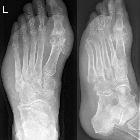
- joints: 1MTP joint most common (known as podagra when it involves this joint); hands and feet are also common
- less common: bones, tendons, bursae
Radiographic features
Most radiographic findings include the skeletal system.
Plain radiograph
Characteristic radiologic changes occur in the chronic stage, though not all patients progress to this. There is a predilection for the small joints of the hands and feet. Chondrocalcinosis is present in ~5%.
Joints
- joint effusion (earliest sign)
- preservation of joint space until late stages of the disease
- an absence of periarticular osteopenia
- eccentric erosions
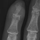
- the typical appearance is the presence of well-defined “punched-out” erosions with sclerotic margins in a marginal and juxta-articular distribution, with overhanging edges (see case 12), also known as rat bite erosions
Bone
- punched-out lytic bone lesions
- overhanging sclerotic margins
- osteonecrosis
- mineralization is normal
Surrounding soft tissues
- tophi: pathognomonic
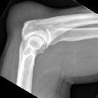
- olecranon and prepatellar bursitis
- periarticular soft tissue swelling due to crystal deposition in tophi around the joints is common
- the soft tissue swelling may be hyperdense due to the crystals, and the tophi can calcify (uncommon in the absence of renal disease)
Ultrasound
While there can be variation in appearance, tophi tend to be hyperechoic, heterogeneous, and have poorly defined contours. They can form multiple groups with surrounding anechoic haloes .
CT
Findings generally reflect those on the plain radiograph.
Dual-energy CT can distinguish between urate mineralization and calcification, which may be useful for cases where the clinical and biochemical presentation is atypical . Allowing for not only visualization and characterization, but also quantification of monosodium urate crystal deposits, it can be used for treatment monitoring as well .
MRI
Signal characteristics of gouty tophi are usually:
- T1: isointense
- T2
- variable
- the majority of lesions are characteristically heterogeneously hypointense
- T1 C+ (Gd): tophus often enhances
Treatment and prognosis
Acutely, gout can be managed with non-steroidal anti-inflammatory drugs (e.g. naproxen), colchicine, prednisolone, or newer cytokine blocking agents (e.g. IL-1 blockers such as anakinra or canakinumab) if refractory disease . In the long-term, xanthine oxidase inhibitors (e.g. allopurinol or feboxustat), uricosuric drugs (e.g. probenecid), or uricase agents (e.g. pegloticase) may be used to reduce urate levels and prevent further acute flares . Tophaceous gout can also be managed with surgical excision of symptomatic lesions .
Siehe auch:
- Aseptische Knochennekrose
- Gelenkerguss
- primärer Hyperparathyreoidismus
- Podagra
- diabetisches Fußsyndrom
- Chondrokalzinose
- Gichtfuß
- Kalziumpyrophosphat-Ablagerungskrankheit
- Kristallarthropathie
- Multi-Energy-Computertomographie
- dual energy CT Gicht
und weiter:
- diffus hypointenses Knochenmarksignal in T1
- Weichteilverkalkungen
- animal and animal produce inspired signs
- tumoröse Kalzinose
- Gichtarthritis am Finger
- Sakroiliitis
- Psoriasisarthritis
- Achillessehnenruptur
- Tendovaginitis
- Quadrizeps-Sehnenruptur
- oberflächliche Weichteilläsionen der Extremitäten
- radiologisches muskuloskelettales Curriculum
- conditions involving skin and bone
- Gefäßverkalkungen
- tendinopathy
- chondrocalcinosis (mnemonic)
- Weichteiltumoren der Extremitäten
- tendon pathology
- differential diagnosis for metatarsal region pain
- Rheuma Differentialdiagnose
- dens erosion
- temporomandibular joint inflammation
- echogenic renal pyramids
- synoviale Raumforderungen
- Arthritis
- Arthritis Knie
- calcium pyrophosphate dihydrate deposition arthropathy
- Weichteilverkalkungen der Hand
- palindromer Rheumatismus
- Gicht MRT
- multi-centric reticulohistiocytosis
- Gicht LWS
- secondary gout
- haemolytic anaemia with secondary gout
- secondary gout in primary myelofibrosis
- Merkspruch Weichteilverkalkungen

 Assoziationen und Differentialdiagnosen zu Gicht:


